1FOE

CRYSTAL STRUCTURE OF RAC1 IN COMPLEX WITH THE GUANINE NUCLEOTIDE EXCHANGE REGION OF TIAM1


Experimental Data Snapshot

  • Method: X-RAY DIFFRACTION
  • Resolution: 2.80 Å
  • R-Value Free: 0.293 
  • R-Value Work: 0.262 

wwPDB Validation   3D Report Full Report


This is version 1.2 of the entry. See complete history


Literature

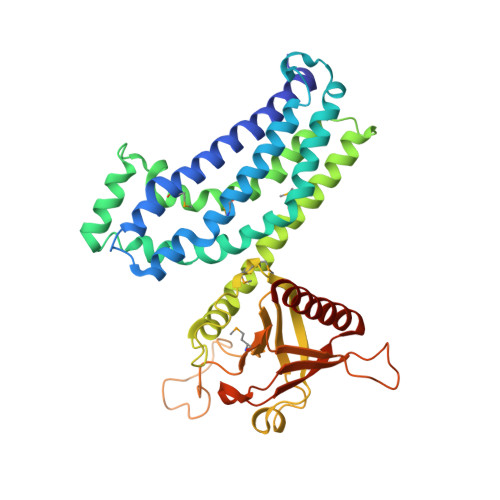
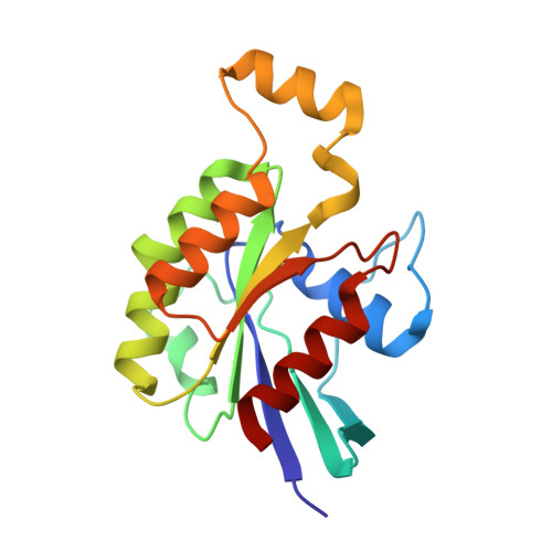
Crystal structure of Rac1 in complex with the guanine nucleotide exchange region of Tiam1.

Worthylake, D.K.Rossman, K.L.Sondek, J.

(2000) Nature 408: 682-688

  • DOI: https://doi.org/10.1038/35047014
  • Primary Citation of Related Structures:  
    1FOE

  • PubMed Abstract: 

    The principal guanine nucleotide exchange factors for Rho family G proteins contain tandem Dbl-homology (DH) and pleckstrin-homology (PH) domains that catalyse nucleotide exchange and the activation of G proteins. Here we have determined the crystal structure of the DH and PH domains of the T-lymphoma invasion and metastasis factor 1 (Tiam1) protein in complex with its cognate Rho family G protein, Rac1. The two switch regions of Rac1 are stabilized in conformations that disrupt both magnesium binding and guanine nucleotide interaction. The resulting cleft in Rac1 is devoid of nucleotide and highly exposed to solvent. The PH domain of Tiam1 does not contact Rac1, and the position and orientation of the PH domain is markedly altered relative to the structure of the uncomplexed, GTPase-free DH/PH element from Sos1. The Tiam1/Rac1 structure highlights the interactions that catalyse nucleotide exchange on Rho family G proteins, and illustrates structural determinants dictating specificity between individual Rho family members and their associated Dbl-related guanine nucleotide exchange factors.


  • Organizational Affiliation

    Department of Pharmacology, Lineberger Comprehensive Cancer Center, The University of North Carolina at Chapel Hill, 27599, USA.


Macromolecules
Find similar proteins by:  (by identity cutoff)  |  3D Structure
Entity ID: 1
MoleculeChains Sequence LengthOrganismDetailsImage
T-LYMPHOMA INVASION AND METASTASIS INDUCING PROTEIN 1
A, C, E, G
377Mus musculusMutation(s): 7 
Membrane Entity: Yes 
UniProt & NIH Common Fund Data Resources
Find proteins for Q60610 (Mus musculus)
Explore Q60610 
Go to UniProtKB:  Q60610
IMPC:  MGI:103306
Entity Groups  
Sequence Clusters30% Identity50% Identity70% Identity90% Identity95% Identity100% Identity
UniProt GroupQ60610
Sequence Annotations
Expand
  • Reference Sequence
Find similar proteins by:  (by identity cutoff)  |  3D Structure
Entity ID: 2
MoleculeChains Sequence LengthOrganismDetailsImage
RAS-RELATED C3 BOTULINUM TOXIN SUBSTRATE
B, D, F, H
177Homo sapiensMutation(s): 0 
Membrane Entity: Yes 
UniProt & NIH Common Fund Data Resources
Find proteins for P63000 (Homo sapiens)
Explore P63000 
Go to UniProtKB:  P63000
PHAROS:  P63000
GTEx:  ENSG00000136238 
Entity Groups  
Sequence Clusters30% Identity50% Identity70% Identity90% Identity95% Identity100% Identity
UniProt GroupP63000
Sequence Annotations
Expand
  • Reference Sequence
Small Molecules
Modified Residues  1 Unique
IDChains TypeFormula2D DiagramParent
MSE
Query on MSE
A, C, E, G
L-PEPTIDE LINKINGC5 H11 N O2 SeMET
Experimental Data & Validation

Experimental Data

  • Method: X-RAY DIFFRACTION
  • Resolution: 2.80 Å
  • R-Value Free: 0.293 
  • R-Value Work: 0.262 
  • Space Group: C 1 2 1
Unit Cell:
Length ( Å )Angle ( ˚ )
a = 186.95α = 90
b = 149.27β = 121
c = 149.21γ = 90
Software Package:
Software NamePurpose
MLPHAREphasing
CNSrefinement
DENZOdata reduction
SCALEPACKdata scaling

Structure Validation

View Full Validation Report


Entry History 

Deposition Data

Revision History  (Full details and data files)

  • Version 1.0: 2001-01-17
    Type: Initial release
  • Version 1.1: 2008-04-27
    Changes: Version format compliance
  • Version 1.2: 2011-07-13
    Changes: Version format compliance