1FRT

CRYSTAL STRUCTURE OF THE COMPLEX OF RAT NEONATAL FC RECEPTOR WITH FC


Experimental Data Snapshot

  • Method: X-RAY DIFFRACTION
  • Resolution: 4.50 Å
  • R-Value Work: 0.423 
  • R-Value Observed: 0.423 

wwPDB Validation   3D Report Full Report


This is version 2.0 of the entry. See complete history


Literature

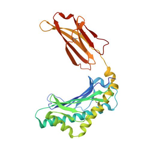
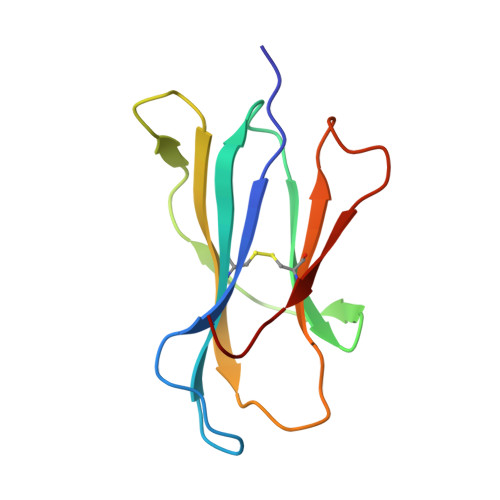
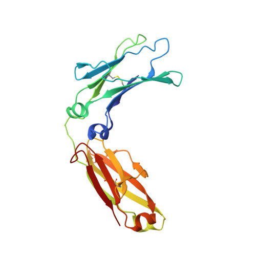
Crystal structure of the complex of rat neonatal Fc receptor with Fc.

Burmeister, W.P.Huber, A.H.Bjorkman, P.J.

(1994) Nature 372: 379-383

  • DOI: https://doi.org/10.1038/372379a0
  • Primary Citation of Related Structures:  
    1FRT

  • PubMed Abstract: 

    The neonatal Fc receptor (FcRn) transports maternal immunoglobulin G (IgG) to the bloodstream of the newborn. FcRn is structurally similar to class I major histocompatibility complex (MHC) molecules, despite differences in the ligands they bind (the Fc portion of IgG and antigenic peptides, respectively). A low-resolution crystal structure of the complex between FcRn and Fc localizes the binding site for Fc to the side of FcRn, distinct from the tops of the alpha 1 and alpha 2 domains which serve as the peptide and T-cell receptor binding sites in class I molecules. FcRn binds to Fc at the interface between the Fc CH2 and CH3 domains, which contains several histidine residues that could account for the sharply pH-dependent FcRn/IgG interaction. A dimer of FcRn heterodimers observed in the co-crystals and in the crystals of FcRn alone could be involved in binding Fc, correlating with the 2:1 binding stoichiometry between FcRn and IgG (ref. 4) and suggesting an unusual orientation of FcRn on the membrane.


  • Organizational Affiliation

    Division of Biology 156-29, California Institute of Technology, Pasadena 91125.


Macromolecules
Find similar proteins by:  (by identity cutoff)  |  3D Structure
Entity ID: 1
MoleculeChains Sequence LengthOrganismDetailsImage
NEONATAL FC RECEPTOR269Rattus norvegicusMutation(s): 0 
Gene Names: BETA-2-MICROGLOBULIN
UniProt
Find proteins for P13599 (Rattus norvegicus)
Explore P13599 
Go to UniProtKB:  P13599
Entity Groups  
Sequence Clusters30% Identity50% Identity70% Identity90% Identity95% Identity100% Identity
UniProt GroupP13599
Sequence Annotations
Expand
  • Reference Sequence
Find similar proteins by:  (by identity cutoff)  |  3D Structure
Entity ID: 2
MoleculeChains Sequence LengthOrganismDetailsImage
BETA 2-MICROGLOBULIN99Rattus norvegicusMutation(s): 0 
Gene Names: BETA-2-MICROGLOBULIN
UniProt
Find proteins for P07151 (Rattus norvegicus)
Explore P07151 
Go to UniProtKB:  P07151
Entity Groups  
Sequence Clusters30% Identity50% Identity70% Identity90% Identity95% Identity100% Identity
UniProt GroupP07151
Sequence Annotations
Expand
  • Reference Sequence
Find similar proteins by:  (by identity cutoff)  |  3D Structure
Entity ID: 3
MoleculeChains Sequence LengthOrganismDetailsImage
IGG FC205Rattus norvegicusMutation(s): 0 
Gene Names: BETA-2-MICROGLOBULIN
Entity Groups  
Sequence Clusters30% Identity50% Identity70% Identity90% Identity95% Identity100% Identity
Sequence Annotations
Expand
  • Reference Sequence
Oligosaccharides

Help

Entity ID: 4
MoleculeChains Length2D Diagram Glycosylation3D Interactions
alpha-L-fucopyranose-(1-6)-2-acetamido-2-deoxy-beta-D-glucopyranose
D
2N-Glycosylation
Glycosylation Resources
GlyTouCan:  G86851RC
GlyCosmos:  G86851RC
GlyGen:  G86851RC
Entity ID: 5
MoleculeChains Length2D Diagram Glycosylation3D Interactions
beta-D-galactopyranose-(1-4)-2-acetamido-2-deoxy-beta-D-glucopyranose-(1-2)-alpha-D-mannopyranose-(1-6)-[2-acetamido-2-deoxy-beta-D-glucopyranose-(1-2)-alpha-D-mannopyranose-(1-3)]beta-D-mannopyranose-(1-4)-2-acetamido-2-deoxy-beta-D-glucopyranose-(1-4)-[alpha-L-fucopyranose-(1-6)]2-acetamido-2-deoxy-beta-D-glucopyranose
E
9N-Glycosylation
Glycosylation Resources
GlyTouCan:  G27919IH
GlyCosmos:  G27919IH
GlyGen:  G27919IH
Small Molecules
Ligands 1 Unique
IDChains Name / Formula / InChI Key2D Diagram3D Interactions
NAG
Query on NAG

Download Ideal Coordinates CCD File 
F [auth A]2-acetamido-2-deoxy-beta-D-glucopyranose
C8 H15 N O6
OVRNDRQMDRJTHS-FMDGEEDCSA-N
Experimental Data & Validation

Experimental Data

  • Method: X-RAY DIFFRACTION
  • Resolution: 4.50 Å
  • R-Value Work: 0.423 
  • R-Value Observed: 0.423 
  • Space Group: I 21 21 21
Unit Cell:
Length ( Å )Angle ( ˚ )
a = 125α = 90
b = 145β = 90
c = 216.5γ = 90
Software Package:
Software NamePurpose
X-PLORmodel building
X-PLORrefinement
X-PLORphasing

Structure Validation

View Full Validation Report


Entry History 

Deposition Data

Revision History  (Full details and data files)

  • Version 1.0: 1995-02-14
    Type: Initial release
  • Version 1.1: 2008-03-03
    Changes: Version format compliance
  • Version 1.2: 2011-07-13
    Changes: Non-polymer description, Version format compliance
  • Version 1.3: 2020-02-05
    Changes: Advisory, Data collection, Database references, Derived calculations, Other
  • Version 2.0: 2020-07-29
    Type: Remediation
    Reason: Carbohydrate remediation
    Changes: Advisory, Atomic model, Data collection, Derived calculations, Structure summary