1MKO

A Fourth Quaternary Structure of Human Hemoglobin A at 2.18 A Resolution


Experimental Data Snapshot

  • Method: X-RAY DIFFRACTION
  • Resolution: 2.18 Å
  • R-Value Free: 0.284 
  • R-Value Work: 0.200 
  • R-Value Observed: 0.200 

wwPDB Validation   3D Report Full Report


Ligand Structure Quality Assessment 


This is version 1.4 of the entry. See complete history


Literature

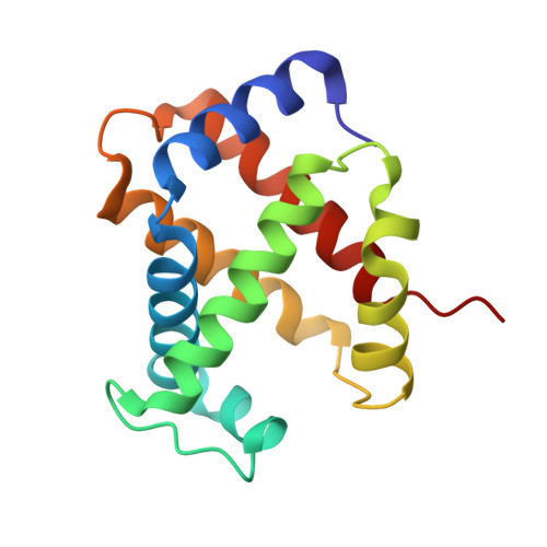
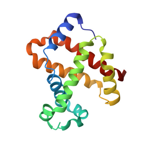
The enigma of the liganded hemoglobin end state: a novel quaternary structure of human carbonmonoxy hemoglobin.

Safo, M.K.Abraham, D.J.

(2005) Biochemistry 44: 8347-8359

  • DOI: https://doi.org/10.1021/bi050412q
  • Primary Citation of Related Structures:  
    1MKO, 1YZI

  • PubMed Abstract: 

    The liganded hemoglobin (Hb) high-salt crystallization condition described by Max Perutz has generated three different crystals of human adult carbonmonoxy hemoglobin (COHbA). The first crystal is isomorphous with the "classical" liganded or R Hb structure. The second crystal reveals a new liganded Hb quaternary structure, RR2, that assumes an intermediate conformation between the R form and another liganded Hb quaternary structure, R2, which was discovered more than a decade ago. Like the R2 structure, the diagnostic R state hydrogen bond between beta2His97 and alpha1Thr38 is missing in the RR2 structure. The third crystal adopts a novel liganded Hb conformation, which we have termed R3, and it shows substantial quaternary structural differences from the R, RR2, and R2 structures. The quaternary structure differences between T and R3 are as large as those between T and R2; however, the T --> R3 and T --> R2 transitions are in different directions as defined by rigid-body screw rotation. Moreover, R3 represents an end state. Compared to all known liganded Hb structures, R3 shows remarkably reduced strain at the alpha-heme, reduced steric contact between the beta-heme ligand and the distal residues, smaller alpha- and beta-clefts, and reduced alpha1-alpha2 and beta1-beta2 iron-iron distances. Together, these unique structural features in R3 should make it the most relaxed and/or greatly enhance its affinity for oxygen compared to the other liganded Hbs. The current Hb structure-function relationships that are now based on T --> R, T -->R --> R2, or T --> R2 --> R transitions may have to be reexamined to take into account the RR2 and R3 liganded structures.


  • Organizational Affiliation

    School of Pharmacy, Department of Medicinal Chemistry, and Institute for Structural Biology and Drug Discovery, Virginia Commonwealth University, Richmond, Virginia 23219, USA. msafo@mail2.vcu.edu


Macromolecules
Find similar proteins by:  (by identity cutoff)  |  3D Structure
Entity ID: 1
MoleculeChains Sequence LengthOrganismDetailsImage
Hemoglobin alpha chain
A, C
141Homo sapiensMutation(s): 0 
UniProt & NIH Common Fund Data Resources
Find proteins for P69905 (Homo sapiens)
Explore P69905 
Go to UniProtKB:  P69905
PHAROS:  P69905
Entity Groups  
Sequence Clusters30% Identity50% Identity70% Identity90% Identity95% Identity100% Identity
UniProt GroupP69905
Sequence Annotations
Expand
  • Reference Sequence
Find similar proteins by:  (by identity cutoff)  |  3D Structure
Entity ID: 2
MoleculeChains Sequence LengthOrganismDetailsImage
Hemoglobin beta chain
B, D
146Homo sapiensMutation(s): 0 
UniProt & NIH Common Fund Data Resources
Find proteins for P68871 (Homo sapiens)
Explore P68871 
Go to UniProtKB:  P68871
PHAROS:  P68871
GTEx:  ENSG00000244734 
Entity Groups  
Sequence Clusters30% Identity50% Identity70% Identity90% Identity95% Identity100% Identity
UniProt GroupP68871
Sequence Annotations
Expand
  • Reference Sequence
Small Molecules
Ligands 3 Unique
IDChains Name / Formula / InChI Key2D Diagram3D Interactions
HEM
Query on HEM

Download Ideal Coordinates CCD File 
G [auth A],
I [auth B],
L [auth C],
O [auth D]
PROTOPORPHYRIN IX CONTAINING FE
C34 H32 Fe N4 O4
KABFMIBPWCXCRK-RGGAHWMASA-L
PO4
Query on PO4

Download Ideal Coordinates CCD File 
E [auth A],
J [auth C],
M [auth D]
PHOSPHATE ION
O4 P
NBIIXXVUZAFLBC-UHFFFAOYSA-K
CMO
Query on CMO

Download Ideal Coordinates CCD File 
F [auth A],
H [auth B],
K [auth C],
N [auth D]
CARBON MONOXIDE
C O
UGFAIRIUMAVXCW-UHFFFAOYSA-N
Experimental Data & Validation

Experimental Data

  • Method: X-RAY DIFFRACTION
  • Resolution: 2.18 Å
  • R-Value Free: 0.284 
  • R-Value Work: 0.200 
  • R-Value Observed: 0.200 
  • Space Group: P 21 21 21
Unit Cell:
Length ( Å )Angle ( ˚ )
a = 65.53α = 90
b = 154.57β = 90
c = 55.27γ = 90
Software Package:
Software NamePurpose
bioteXdata collection
bioteXdata reduction
AMoREphasing
CNSrefinement
bioteXdata scaling

Structure Validation

View Full Validation Report


Ligand Structure Quality Assessment 


Entry History 

Deposition Data

Revision History  (Full details and data files)

  • Version 1.0: 2002-09-06
    Type: Initial release
  • Version 1.1: 2008-04-28
    Changes: Version format compliance
  • Version 1.2: 2011-07-13
    Changes: Version format compliance
  • Version 1.3: 2017-10-11
    Changes: Data collection, Refinement description
  • Version 1.4: 2024-02-14
    Changes: Data collection, Database references, Derived calculations, Refinement description