1STY

THE ALPHA ANEURISM: A STRUCTURAL MOTIF REVEALED IN AN INSERTION MUTANT OF STAPHYLOCOCCAL NUCLEASE


Experimental Data Snapshot

  • Method: X-RAY DIFFRACTION
  • Resolution: 1.67 Å
  • R-Value Work: 0.170 

wwPDB Validation   3D Report Full Report


This is version 1.6 of the entry. See complete history


Literature

The alpha aneurism: a structural motif revealed in an insertion mutant of staphylococcal nuclease.

Keefe, L.J.Sondek, J.Shortle, D.Lattman, E.E.

(1993) Proc Natl Acad Sci U S A 90: 3275-3279

  • DOI: https://doi.org/10.1073/pnas.90.8.3275
  • Primary Citation of Related Structures:  
    1STY

  • PubMed Abstract: 

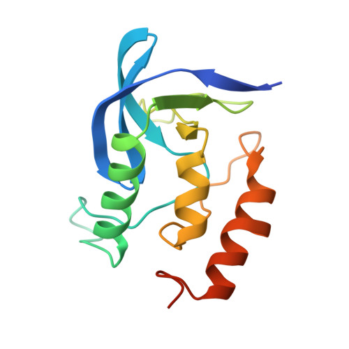
    The x-ray crystal structure of a mutant of staphylococcal nuclease that contains a single glycine residue inserted in the C-terminal alpha-helix has been solved to 1.67 A resolution and refined to a crystallographic R value of 0.170. This inserted glycine residue is accommodated in the alpha-helix by formation of a previously uncharacterized bulge, which we term the alpha aneurism. A conformational search of known protein structures has identified the alpha aneurism in a number of protein families, including the histocompatibility antigens and hemoglobins.


  • Organizational Affiliation

    Department of Biophysics and Biophysical Chemistry, Johns Hopkins University School of Medicine, Baltimore, MD 21205-2185.


Macromolecules
Find similar proteins by:  (by identity cutoff)  |  3D Structure
Entity ID: 1
MoleculeChains Sequence LengthOrganismDetailsImage
STAPHYLOCOCCAL NUCLEASE150Staphylococcus aureusMutation(s): 0 
EC: 3.1.31.1
UniProt
Find proteins for P00644 (Staphylococcus aureus)
Explore P00644 
Go to UniProtKB:  P00644
Entity Groups  
Sequence Clusters30% Identity50% Identity70% Identity90% Identity95% Identity100% Identity
UniProt GroupP00644
Sequence Annotations
Expand
  • Reference Sequence
Small Molecules
Ligands 2 Unique
IDChains Name / Formula / InChI Key2D Diagram3D Interactions
THP
Query on THP

Download Ideal Coordinates CCD File 
C [auth A]THYMIDINE-3',5'-DIPHOSPHATE
C10 H16 N2 O11 P2
CSNCBOPUCJOHLS-XLPZGREQSA-N
CA
Query on CA

Download Ideal Coordinates CCD File 
B [auth A]CALCIUM ION
Ca
BHPQYMZQTOCNFJ-UHFFFAOYSA-N
Experimental Data & Validation

Experimental Data

  • Method: X-RAY DIFFRACTION
  • Resolution: 1.67 Å
  • R-Value Work: 0.170 
  • Space Group: P 41
Unit Cell:
Length ( Å )Angle ( ˚ )
a = 46.952α = 90
b = 46.952β = 90
c = 62.895γ = 90
Software Package:
Software NamePurpose
X-PLORmodel building
PROLSQrefinement
X-PLORrefinement
X-PLORphasing

Structure Validation

View Full Validation Report


Entry History 

Deposition Data

Revision History  (Full details and data files)

  • Version 1.0: 1993-04-15
    Type: Initial release
  • Version 1.1: 2008-03-03
    Changes: Version format compliance
  • Version 1.2: 2011-07-13
    Changes: Version format compliance
  • Version 1.3: 2017-11-29
    Changes: Derived calculations, Other
  • Version 1.4: 2019-07-17
    Changes: Data collection, Refinement description
  • Version 1.5: 2019-08-14
    Changes: Data collection, Refinement description
  • Version 1.6: 2024-02-14
    Changes: Data collection, Database references, Derived calculations