1T89

CRYSTAL STRUCTURE OF A HUMAN TYPE III FC GAMMA RECEPTOR IN COMPLEX WITH AN FC FRAGMENT OF IGG1 (HEXAGONAL)


Experimental Data Snapshot

  • Method: X-RAY DIFFRACTION
  • Resolution: 3.50 Å
  • R-Value Free: 0.309 
  • R-Value Work: 0.247 

wwPDB Validation   3D Report Full Report


This is version 2.1 of the entry. See complete history


Literature

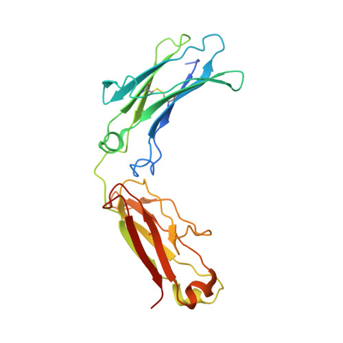
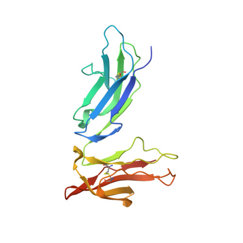
The structure of a human type III Fcgamma receptor in complex with Fc

Radaev, S.Motyka, S.Fridman, W.-H.Sautes-Fridman, C.Sun, P.D.

(2001) J Biol Chem 276: 16469-16477

  • DOI: https://doi.org/10.1074/jbc.M100350200
  • Primary Citation of Related Structures:  
    1T83, 1T89

  • PubMed Abstract: 

    Fcgamma receptors mediate antibody-dependent inflammatory responses and cytotoxicity as well as certain autoimmune dysfunctions. Here we report the crystal structure of a human Fc receptor (FcgammaRIIIB) in complex with an Fc fragment of human IgG1 determined from orthorhombic and hexagonal crystal forms at 3.0- and 3.5-A resolution, respectively. The refined structures from the two crystal forms are nearly identical with no significant discrepancies between the coordinates. Regions of the C-terminal domain of FcgammaRIII, including the BC, C'E, FG loops, and the C' beta-strand, bind asymmetrically to the lower hinge region, residues Leu(234)-Pro(238), of both Fc chains creating a 1:1 receptor-ligand stoichiometry. Minor conformational changes are observed in both the receptor and Fc upon complex formation. Hydrophobic residues, hydrogen bonds, and salt bridges are distributed throughout the receptor.Fc interface. Sequence comparisons of the receptor-ligand interface residues suggest a conserved binding mode common to all members of immunoglobulin-like Fc receptors. Structural comparison between FcgammaRIII.Fc and FcepsilonRI.Fc complexes highlights the differences in ligand recognition between the high and low affinity receptors. Although not in direct contact with the receptor, the carbohydrate attached to the conserved glycosylation residue Asn(297) on Fc may stabilize the conformation of the receptor-binding epitope on Fc. An antibody-FcgammaRIII model suggests two possible ligand-induced receptor aggregations.


  • Organizational Affiliation

    Structural Biology Section, Laboratory of Immunogenetics, NIAID, National Institutes of Health, Rockville, Maryland 20852, USA.


Macromolecules
Find similar proteins by:  (by identity cutoff)  |  3D Structure
Entity ID: 1
MoleculeChains Sequence LengthOrganismDetailsImage
recombinant IgG1 heavy chain
A, B
224Homo sapiensMutation(s): 0 
UniProt & NIH Common Fund Data Resources
Find proteins for P01857 (Homo sapiens)
Explore P01857 
Go to UniProtKB:  P01857
PHAROS:  P01857
GTEx:  ENSG00000211896 
Entity Groups  
Sequence Clusters30% Identity50% Identity70% Identity90% Identity95% Identity100% Identity
UniProt GroupP01857
Sequence Annotations
Expand
  • Reference Sequence
Find similar proteins by:  (by identity cutoff)  |  3D Structure
Entity ID: 2
MoleculeChains Sequence LengthOrganismDetailsImage
Low affinity immunoglobulin gamma Fc region receptor III-B176Homo sapiensMutation(s): 0 
UniProt & NIH Common Fund Data Resources
Find proteins for O75015 (Homo sapiens)
Explore O75015 
Go to UniProtKB:  O75015
PHAROS:  O75015
GTEx:  ENSG00000162747 
Entity Groups  
Sequence Clusters30% Identity50% Identity70% Identity90% Identity95% Identity100% Identity
UniProt GroupO75015
Sequence Annotations
Expand
  • Reference Sequence
Oligosaccharides

Help

Entity ID: 3
MoleculeChains Length2D Diagram Glycosylation3D Interactions
beta-D-galactopyranose-(1-4)-2-acetamido-2-deoxy-alpha-D-glucopyranose-(1-2)-alpha-D-mannopyranose-(1-6)-[beta-D-mannopyranose-(1-3)]beta-D-mannopyranose-(1-4)-2-acetamido-2-deoxy-beta-D-glucopyranose-(1-4)-[beta-L-fucopyranose-(1-6)]2-acetamido-2-deoxy-beta-D-glucopyranose
D
8N-Glycosylation
Glycosylation Resources
GlyTouCan:  G71516NY
GlyCosmos:  G71516NY
GlyGen:  G71516NY
Entity ID: 4
MoleculeChains Length2D Diagram Glycosylation3D Interactions
2-acetamido-2-deoxy-beta-D-glucopyranose-(1-2)-alpha-D-mannopyranose-(1-3)-[2-acetamido-2-deoxy-beta-D-glucopyranose-(1-2)-alpha-D-mannopyranose-(1-6)]beta-D-mannopyranose-(1-4)-2-acetamido-2-deoxy-alpha-D-glucopyranose-(1-4)-[alpha-L-fucopyranose-(1-6)]2-acetamido-2-deoxy-beta-D-glucopyranose
E
8N-Glycosylation
Glycosylation Resources
GlyTouCan:  G01257PC
GlyCosmos:  G01257PC
GlyGen:  G01257PC
Experimental Data & Validation

Experimental Data

  • Method: X-RAY DIFFRACTION
  • Resolution: 3.50 Å
  • R-Value Free: 0.309 
  • R-Value Work: 0.247 
  • Space Group: P 65 2 2
Unit Cell:
Length ( Å )Angle ( ˚ )
a = 114.984α = 90
b = 114.984β = 90
c = 301.4γ = 120
Software Package:
Software NamePurpose
SCALEPACKdata scaling
AMoREphasing
CNSrefinement

Structure Validation

View Full Validation Report


Entry History 

Deposition Data

Revision History  (Full details and data files)

  • Version 1.0: 2004-09-14
    Type: Initial release
  • Version 1.1: 2008-04-30
    Changes: Version format compliance
  • Version 1.2: 2011-07-13
    Changes: Non-polymer description, Version format compliance
  • Version 2.0: 2020-07-29
    Type: Remediation
    Reason: Carbohydrate remediation
    Changes: Advisory, Atomic model, Data collection, Derived calculations, Structure summary
  • Version 2.1: 2023-08-23
    Changes: Advisory, Data collection, Database references, Refinement description, Structure summary