256L

BACTERIOPHAGE T4 LYSOZYME


Experimental Data Snapshot

  • Method: X-RAY DIFFRACTION
  • Resolution: 1.80 Å
  • R-Value Work: 0.163 

wwPDB Validation   3D Report Full Report


This is version 1.5 of the entry. See complete history


Literature

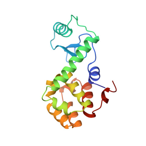
A mutant T4 lysozyme displays five different crystal conformations.

Faber, H.R.Matthews, B.W.

(1990) Nature 348: 263-266

  • DOI: https://doi.org/10.1038/348263a0
  • Primary Citation of Related Structures:  
    256L

  • PubMed Abstract: 

    Phage T4 lysozyme consists of two domains between which is formed the active-site cleft of the enzyme. The crystallographically determined thermal displacement parameters for the protein suggested that the amino terminal of the two domains undergoes 'hinge-bending' motion about an axis passing through the waist of the molecule. Such conformational mobility may be important in allowing access of substrates to the active site of the enzyme. We report here a crystallographic study of a mutant T4 lysozyme which demonstrates further the conformational flexibility of the protein. A mutant form of the enzyme with a methionine residue (Met 6) replaced by isoleucine crystallizes with four independent molecules in the crystal lattice. These four molecules have distinctly different conformations. The mutant protein can also crystallize in standard form with a structure very similar to the wild-type protein. Thus the mutant protein can adopt five different crystal conformations. The isoleucine for methionine substitution at the intersection of the two domains of T4 lysozyme apparently enhances the hinge-bending motion presumed to occur in the wild-type protein, without significantly affecting the catalytic activity or thermal stability of the protein.


  • Organizational Affiliation

    Department of Physics, University of Oregon, Eugene 97403.


Macromolecules
Find similar proteins by:  (by identity cutoff)  |  3D Structure
Entity ID: 1
MoleculeChains Sequence LengthOrganismDetailsImage
LYSOZYME164Tequatrovirus T4Mutation(s): 1 
EC: 3.2.1.17
UniProt
Find proteins for P00720 (Enterobacteria phage T4)
Explore P00720 
Go to UniProtKB:  P00720
Entity Groups  
Sequence Clusters30% Identity50% Identity70% Identity90% Identity95% Identity100% Identity
UniProt GroupP00720
Sequence Annotations
Expand
  • Reference Sequence
Experimental Data & Validation

Experimental Data

  • Method: X-RAY DIFFRACTION
  • Resolution: 1.80 Å
  • R-Value Work: 0.163 
  • Space Group: P 32 2 1
Unit Cell:
Length ( Å )Angle ( ˚ )
a = 61.1α = 90
b = 61.1β = 90
c = 97.6γ = 120
Software Package:
Software NamePurpose
TNTrefinement
ROSSMANNdata reduction
CAMBRIDGE/ODPROCdata scaling
CCP4data scaling
TNTphasing

Structure Validation

View Full Validation Report


Entry History 

Deposition Data

Revision History  (Full details and data files)

  • Version 1.0: 1998-05-27
    Type: Initial release
  • Version 1.1: 2008-03-24
    Changes: Version format compliance
  • Version 1.2: 2011-07-13
    Changes: Version format compliance
  • Version 1.3: 2021-11-03
    Changes: Database references
  • Version 1.4: 2024-02-14
    Changes: Data collection
  • Version 1.5: 2024-04-03
    Changes: Refinement description