2B3P

Crystal structure of a superfolder green fluorescent protein


Experimental Data Snapshot

  • Method: X-RAY DIFFRACTION
  • Resolution: 1.40 Å
  • R-Value Free: 0.218 
  • R-Value Work: 0.206 
  • R-Value Observed: 0.206 

wwPDB Validation   3D Report Full Report


This is version 1.5 of the entry. See complete history


Literature

Engineering and characterization of a superfolder green fluorescent protein.

Pedelacq, J.D.Cabantous, S.Tran, T.Terwilliger, T.C.Waldo, G.S.

(2006) Nat Biotechnol 24: 79-88

  • DOI: https://doi.org/10.1038/nbt1172
  • Primary Citation of Related Structures:  
    2B3P, 2B3Q

  • PubMed Abstract: 

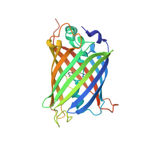
    Existing variants of green fluorescent protein (GFP) often misfold when expressed as fusions with other proteins. We have generated a robustly folded version of GFP, called 'superfolder' GFP, that folds well even when fused to poorly folded polypeptides. Compared to 'folding reporter' GFP, a folding-enhanced GFP containing the 'cycle-3' mutations and the 'enhanced GFP' mutations F64L and S65T, superfolder GFP shows improved tolerance of circular permutation, greater resistance to chemical denaturants and improved folding kinetics. The fluorescence of Escherichia coli cells expressing each of eighteen proteins from Pyrobaculum aerophilum as fusions with superfolder GFP was proportional to total protein expression. In contrast, fluorescence of folding reporter GFP fusion proteins was strongly correlated with the productive folding yield of the passenger protein. X-ray crystallographic structural analyses helped explain the enhanced folding of superfolder GFP relative to folding reporter GFP.


  • Organizational Affiliation

    Bioscience Division, MS-M888, Los Alamos National Laboratory, Los Alamos, New Mexico 87545, USA.


Macromolecules
Find similar proteins by:  (by identity cutoff)  |  3D Structure
Entity ID: 1
MoleculeChains Sequence LengthOrganismDetailsImage
green fluorescent protein244Aequorea victoriaMutation(s): 12 
Gene Names: gft
UniProt
Find proteins for P42212 (Aequorea victoria)
Explore P42212 
Go to UniProtKB:  P42212
Entity Groups  
Sequence Clusters30% Identity50% Identity70% Identity90% Identity95% Identity100% Identity
UniProt GroupP42212
Sequence Annotations
Expand
  • Reference Sequence
Small Molecules
Ligands 2 Unique
IDChains Name / Formula / InChI Key2D Diagram3D Interactions
CD
Query on CD

Download Ideal Coordinates CCD File 
B [auth A]
C [auth A]
D [auth A]
E [auth A]
F [auth A]
B [auth A],
C [auth A],
D [auth A],
E [auth A],
F [auth A],
G [auth A],
H [auth A],
I [auth A],
J [auth A]
CADMIUM ION
Cd
WLZRMCYVCSSEQC-UHFFFAOYSA-N
ACY
Query on ACY

Download Ideal Coordinates CCD File 
K [auth A]
L [auth A]
M [auth A]
N [auth A]
O [auth A]
K [auth A],
L [auth A],
M [auth A],
N [auth A],
O [auth A],
P [auth A]
ACETIC ACID
C2 H4 O2
QTBSBXVTEAMEQO-UHFFFAOYSA-N
Modified Residues  1 Unique
IDChains TypeFormula2D DiagramParent
CRO
Query on CRO
A
L-PEPTIDE LINKINGC15 H17 N3 O5THR, TYR, GLY
Experimental Data & Validation

Experimental Data

  • Method: X-RAY DIFFRACTION
  • Resolution: 1.40 Å
  • R-Value Free: 0.218 
  • R-Value Work: 0.206 
  • R-Value Observed: 0.206 
  • Space Group: P 31 2 1
Unit Cell:
Length ( Å )Angle ( ˚ )
a = 88.456α = 90
b = 88.456β = 90
c = 69.346γ = 120
Software Package:
Software NamePurpose
CNSrefinement
MOSFLMdata reduction
CCP4data scaling
CNSphasing

Structure Validation

View Full Validation Report


Entry History 

Deposition Data

Revision History  (Full details and data files)

  • Version 1.0: 2005-11-08
    Type: Initial release
  • Version 1.1: 2008-05-01
    Changes: Version format compliance
  • Version 1.2: 2011-07-13
    Changes: Version format compliance
  • Version 1.3: 2021-10-20
    Changes: Advisory, Database references, Derived calculations
  • Version 1.4: 2023-08-23
    Changes: Data collection, Refinement description
  • Version 1.5: 2023-11-15
    Changes: Data collection