2JIZ

The Structure of F1-ATPase inhibited by resveratrol.


Experimental Data Snapshot

  • Method: X-RAY DIFFRACTION
  • Resolution: 2.30 Å
  • R-Value Free: 0.217 
  • R-Value Work: 0.160 
  • R-Value Observed: 0.163 

wwPDB Validation   3D Report Full Report


This is version 1.2 of the entry. See complete history


Literature

Mechanism of Inhibition of Bovine F1-ATPase by Resveratrol and Related Polyphenols.

Gledhill, J.R.Montgomery, M.G.Leslie, A.G.W.Walker, J.E.

(2007) Proc Natl Acad Sci U S A 104: 13632

  • DOI: https://doi.org/10.1073/pnas.0706290104
  • Primary Citation of Related Structures:  
    2JIZ, 2JJ1, 2JJ2

  • PubMed Abstract: 

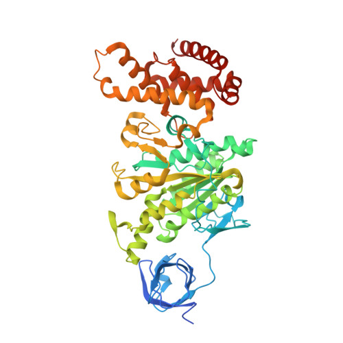
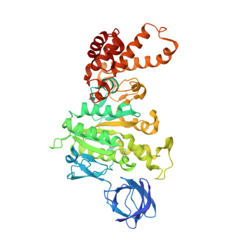
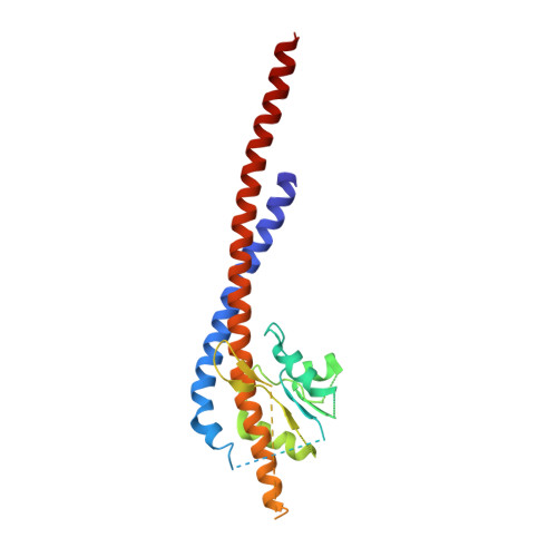
    The structures of F(1)-ATPase from bovine heart mitochondria inhibited with the dietary phytopolyphenol, resveratrol, and with the related polyphenols quercetin and piceatannol have been determined at 2.3-, 2.4- and 2.7-A resolution, respectively. The inhibitors bind to a common site in the inside surface of an annulus made from loops in the three alpha- and three beta-subunits beneath the "crown" of beta-strands in their N-terminal domains. This region of F(1)-ATPase forms a bearing to allow the rotation of the tip of the gamma-subunit inside the annulus during catalysis. The binding site is a hydrophobic pocket between the C-terminal tip of the gamma-subunit and the beta(TP) subunit, and the inhibitors are bound via H-bonds mostly to their hydroxyl moieties mediated by bound water molecules and by hydrophobic interactions. There are no equivalent sites between the gamma-subunit and either the beta(DP) or the beta(E) subunit. The inhibitors probably prevent both the synthetic and hydrolytic activities of the enzyme by blocking both senses of rotation of the gamma-subunit. The beneficial effects of dietary resveratrol may derive in part by preventing mitochondrial ATP synthesis in tumor cells, thereby inducing apoptosis.


  • Organizational Affiliation

    Medical Research Council Dunn Human Nutrition Unit, Wellcome Trust/MRC Building, Hills Road, Cambridge CB2 0XY, United Kingdom.


Macromolecules
Find similar proteins by:  (by identity cutoff)  |  3D Structure
Entity ID: 1
MoleculeChains Sequence LengthOrganismDetailsImage
ATP SYNTHASE SUBUNIT ALPHA HEART ISOFORM
A, B, C, H, I
A, B, C, H, I, J
510Bos taurusMutation(s): 0 
UniProt
Find proteins for P19483 (Bos taurus)
Explore P19483 
Go to UniProtKB:  P19483
Entity Groups  
Sequence Clusters30% Identity50% Identity70% Identity90% Identity95% Identity100% Identity
UniProt GroupP19483
Sequence Annotations
Expand
  • Reference Sequence
Find similar proteins by:  (by identity cutoff)  |  3D Structure
Entity ID: 2
MoleculeChains Sequence LengthOrganismDetailsImage
ATP SYNTHASE SUBUNIT BETA
D, E, F, K, L
D, E, F, K, L, M
482Bos taurusMutation(s): 0 
UniProt
Find proteins for P00829 (Bos taurus)
Explore P00829 
Go to UniProtKB:  P00829
Entity Groups  
Sequence Clusters30% Identity50% Identity70% Identity90% Identity95% Identity100% Identity
UniProt GroupP00829
Sequence Annotations
Expand
  • Reference Sequence
Find similar proteins by:  (by identity cutoff)  |  3D Structure
Entity ID: 3
MoleculeChains Sequence LengthOrganismDetailsImage
ATP SYNTHASE GAMMA CHAIN
G, N
272Bos taurusMutation(s): 0 
UniProt
Find proteins for P05631 (Bos taurus)
Explore P05631 
Go to UniProtKB:  P05631
Entity Groups  
Sequence Clusters30% Identity50% Identity70% Identity90% Identity95% Identity100% Identity
UniProt GroupP05631
Sequence Annotations
Expand
  • Reference Sequence
Small Molecules
Ligands 7 Unique
IDChains Name / Formula / InChI Key2D Diagram3D Interactions
ANP
Query on ANP

Download Ideal Coordinates CCD File 
EA [auth F]
HA [auth H]
LA [auth I]
O [auth A]
OA [auth J]
EA [auth F],
HA [auth H],
LA [auth I],
O [auth A],
OA [auth J],
S [auth B],
W [auth C],
XA [auth M]
PHOSPHOAMINOPHOSPHONIC ACID-ADENYLATE ESTER
C10 H17 N6 O12 P3
PVKSNHVPLWYQGJ-KQYNXXCUSA-N
ADP
Query on ADP

Download Ideal Coordinates CCD File 
RA [auth K],
Z [auth D]
ADENOSINE-5'-DIPHOSPHATE
C10 H15 N5 O10 P2
XTWYTFMLZFPYCI-KQYNXXCUSA-N
STL
Query on STL

Download Ideal Coordinates CCD File 
GA [auth G],
ZA [auth N]
RESVERATROL
C14 H12 O3
LUKBXSAWLPMMSZ-OWOJBTEDSA-N
PO4
Query on PO4

Download Ideal Coordinates CCD File 
DA [auth E],
WA [auth L]
PHOSPHATE ION
O4 P
NBIIXXVUZAFLBC-UHFFFAOYSA-K
GOL
Query on GOL

Download Ideal Coordinates CCD File 
CA [auth D]
JA [auth H]
KA [auth H]
NA [auth I]
Q [auth A]
CA [auth D],
JA [auth H],
KA [auth H],
NA [auth I],
Q [auth A],
QA [auth J],
R [auth A],
U [auth B],
UA [auth K],
V [auth B],
VA [auth K],
Y [auth C]
GLYCEROL
C3 H8 O3
PEDCQBHIVMGVHV-UHFFFAOYSA-N
AZI
Query on AZI

Download Ideal Coordinates CCD File 
BA [auth D],
TA [auth K]
AZIDE ION
N3
IVRMZWNICZWHMI-UHFFFAOYSA-N
MG
Query on MG

Download Ideal Coordinates CCD File 
AA [auth D]
FA [auth F]
IA [auth H]
MA [auth I]
P [auth A]
AA [auth D],
FA [auth F],
IA [auth H],
MA [auth I],
P [auth A],
PA [auth J],
SA [auth K],
T [auth B],
X [auth C],
YA [auth M]
MAGNESIUM ION
Mg
JLVVSXFLKOJNIY-UHFFFAOYSA-N
Experimental Data & Validation

Experimental Data

  • Method: X-RAY DIFFRACTION
  • Resolution: 2.30 Å
  • R-Value Free: 0.217 
  • R-Value Work: 0.160 
  • R-Value Observed: 0.163 
  • Space Group: P 1 21 1
Unit Cell:
Length ( Å )Angle ( ˚ )
a = 106.776α = 90
b = 277.368β = 90.19
c = 137.827γ = 90
Software Package:
Software NamePurpose
REFMACrefinement
MOSFLMdata reduction
CCP4data scaling
AMoREphasing

Structure Validation

View Full Validation Report


Entry History 

Deposition Data

Revision History  (Full details and data files)

  • Version 1.0: 2007-08-21
    Type: Initial release
  • Version 1.1: 2011-07-13
    Changes: Advisory, Refinement description, Version format compliance
  • Version 1.2: 2023-12-13
    Changes: Data collection, Database references, Derived calculations, Other, Refinement description