2Q59

Crystal Structure of PPARgamma LBD bound to full agonist MRL20


Experimental Data Snapshot

  • Method: X-RAY DIFFRACTION
  • Resolution: 2.20 Å
  • R-Value Free: 0.230 
  • R-Value Work: 0.183 
  • R-Value Observed: 0.185 

wwPDB Validation   3D Report Full Report


Ligand Structure Quality Assessment 


This is version 1.3 of the entry. See complete history


Literature

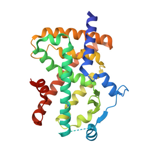
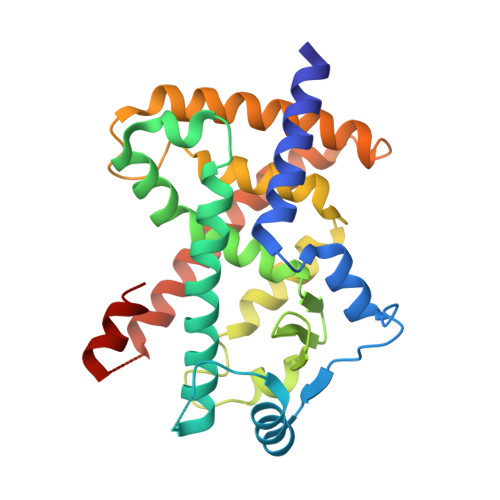
Partial Agonists Activate PPARgamma Using a Helix 12 Independent Mechanism

Bruning, J.B.Chalmers, M.J.Prasad, S.Busby, S.A.Kamenecka, T.M.He, Y.Nettles, K.W.Griffin, P.R.

(2007) Structure 15: 1258-1271

  • DOI: https://doi.org/10.1016/j.str.2007.07.014
  • Primary Citation of Related Structures:  
    2Q59, 2Q5P, 2Q5S, 2Q61, 2Q6R, 2Q6S

  • PubMed Abstract: 

    Binding to helix 12 of the ligand-binding domain of PPARgamma is required for full agonist activity. Previously, the degree of stabilization of the activation function 2 (AF-2) surface was thought to correlate with the degree of agonism and transactivation. To examine this mechanism, we probed structural dynamics of PPARgamma with agonists that induced graded transcriptional responses. Here we present crystal structures and amide H/D exchange (HDX) kinetics for six of these complexes. Amide HDX revealed each ligand induced unique changes to the dynamics of the ligand-binding domain (LBD). Full agonists stabilized helix 12, whereas intermediate and partial agonists did not at all, and rather differentially stabilized other regions of the binding pocket. The gradient of PPARgamma transactivation cannot be accounted for solely through changes to the dynamics of AF-2. Thus, our understanding of allosteric signaling must be extended beyond the idea of a dynamic helix 12 acting as a molecular switch.


  • Organizational Affiliation

    Department of Cancer Biology, The Scripps Research Institute, 5353 Parkside Drive, Jupiter, FL 33458, USA.


Macromolecules
Find similar proteins by:  (by identity cutoff)  |  3D Structure
Entity ID: 1
MoleculeChains Sequence LengthOrganismDetailsImage
Peroxisome Proliferator-Activated Receptor gamma274Homo sapiensMutation(s): 1 
Gene Names: PPARGNR1C3
UniProt & NIH Common Fund Data Resources
Find proteins for P37231 (Homo sapiens)
Explore P37231 
Go to UniProtKB:  P37231
PHAROS:  P37231
GTEx:  ENSG00000132170 
Entity Groups  
Sequence Clusters30% Identity50% Identity70% Identity90% Identity95% Identity100% Identity
UniProt GroupP37231
Sequence Annotations
Expand
  • Reference Sequence
Find similar proteins by:  (by identity cutoff)  |  3D Structure
Entity ID: 2
MoleculeChains Sequence LengthOrganismDetailsImage
Peroxisome Proliferator-Activated Receptor gamma274Homo sapiensMutation(s): 0 
Gene Names: PPARGNR1C3
UniProt & NIH Common Fund Data Resources
Find proteins for P37231 (Homo sapiens)
Explore P37231 
Go to UniProtKB:  P37231
PHAROS:  P37231
GTEx:  ENSG00000132170 
Entity Groups  
Sequence Clusters30% Identity50% Identity70% Identity90% Identity95% Identity100% Identity
UniProt GroupP37231
Sequence Annotations
Expand
  • Reference Sequence
Small Molecules
Ligands 1 Unique
IDChains Name / Formula / InChI Key2D Diagram3D Interactions
240
Query on 240

Download Ideal Coordinates CCD File 
C [auth A],
D [auth B]
(2S)-2-(2-{[1-(4-METHOXYBENZOYL)-2-METHYL-5-(TRIFLUOROMETHOXY)-1H-INDOL-3-YL]METHYL}PHENOXY)PROPANOIC ACID
C28 H24 F3 N O6
BWTOBMCYVACNJZ-KRWDZBQOSA-N
Modified Residues  1 Unique
IDChains TypeFormula2D DiagramParent
CME
Query on CME
A
L-PEPTIDE LINKINGC5 H11 N O3 S2CYS
Binding Affinity Annotations 
IDSourceBinding Affinity
240 BindingDB:  2Q59 IC50: min: 1, max: 2 (nM) from 2 assay(s)
EC50: 2 (nM) from 1 assay(s)
Experimental Data & Validation

Experimental Data

  • Method: X-RAY DIFFRACTION
  • Resolution: 2.20 Å
  • R-Value Free: 0.230 
  • R-Value Work: 0.183 
  • R-Value Observed: 0.185 
  • Space Group: C 1 2 1
Unit Cell:
Length ( Å )Angle ( ˚ )
a = 89.161α = 90
b = 63.939β = 103.43
c = 119.187γ = 90
Software Package:
Software NamePurpose
DENZOdata reduction
SCALEPACKdata scaling
REFMACrefinement
PDB_EXTRACTdata extraction
HKL-2000data collection
HKL-2000data reduction
MOLREPphasing

Structure Validation

View Full Validation Report


Ligand Structure Quality Assessment 


Entry History 

Deposition Data

Revision History  (Full details and data files)

  • Version 1.0: 2007-10-23
    Type: Initial release
  • Version 1.1: 2011-07-13
    Changes: Advisory, Version format compliance
  • Version 1.2: 2017-10-18
    Changes: Refinement description
  • Version 1.3: 2023-08-30
    Changes: Data collection, Database references, Derived calculations, Refinement description