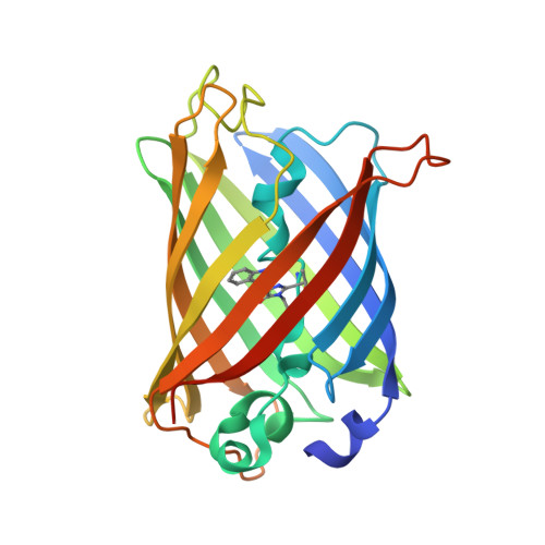
Intrinsic Dynamics in Ecfp and Cerulean Control Fluorescence Quantum Yield.
Lelimousin, M., Noirclerc-Savoye, M., Lazareno-Saez, C., Paetzold, B., Le Vot, S., Chazal, R., Macheboeuf, P., Field, M.J., Bourgeois, D., Royant, A.(2009) Biochemistry 48: 10038
- PubMed: 19754158
- DOI: https://doi.org/10.1021/bi901093w
- Primary Citation of Related Structures:
2WSN, 2WSO - PubMed Abstract:
Enhanced cyan fluorescent protein (ECFP) and its variant Cerulean are genetically encoded fluorophores widely used as donors in FRET-based cell imaging experiments. First, we have confirmed through denaturation experiments that the double-peak spectroscopic signature of these fluorescent proteins originates from the indole ring of the chromophore. Then, to explain the improvement in the fluorescence properties of Cerulean compared to those of ECFP, we have determined the high-resolution crystal structures of these two proteins at physiological pH and performed molecular dynamics simulations. In both proteins, the N-terminal half of the seventh strand exhibits two conformations. These conformations both have a complex set of van der Waals interactions with the chromophore and, as our simulations suggest, they interconvert on a nanosecond time scale. The Y145A and H148D mutations in Cerulean stabilize these interactions and allow the chromophore to be more planar, better packed, and less prone to collisional quenching, albeit only intermittently. As a consequence, the probability of nonradiative decay is significantly decreased. Our results highlight the considerable dynamical flexibility that exists in the vicinity of the tryptophan-based chromophore of these engineered fluorescent proteins and provide insights that should allow the design of mutants with enhanced optical properties.
Organizational Affiliation:
Institut de Biologie Structurale Jean-Pierre Ebel, UMR 5075 CNRS-CEA-Universite Joseph Fourier, F-38027 Grenoble Cedex 1, France.