2X7C

Crystal structure of human kinesin Eg5 in complex with (S)-enastron


Experimental Data Snapshot

  • Method: X-RAY DIFFRACTION
  • Resolution: 1.90 Å
  • R-Value Free: 0.232 
  • R-Value Work: 0.163 
  • R-Value Observed: 0.167 

wwPDB Validation   3D Report Full Report


Ligand Structure Quality Assessment 


This is version 1.2 of the entry. See complete history


Literature

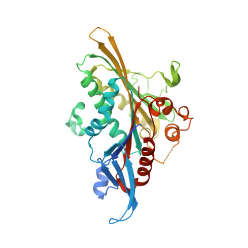
Structural Basis for Inhibition of Eg5 by Dihydropyrimidines: Stereoselectivity of Antimitotic Inhibitors Enastron, Dimethylenastron and Fluorastrol.

Kaan, H.Y.K.Ulaganathan, V.Rath, O.Prokopcova, H.Dallinger, D.Kappe, C.O.Kozielski, F.

(2010) J Med Chem 53: 5676

  • DOI: https://doi.org/10.1021/jm100421n
  • Primary Citation of Related Structures:  
    2X7C, 2X7D, 2X7E

  • PubMed Abstract: 

    Human kinesin Eg5, which plays an essential role in mitosis by establishing the bipolar spindle, has proven to be an interesting drug target for the development of cancer chemotherapeutics. Here, we report the crystal structures of the Eg5 motor domain complexed with enastron, dimethylenastron, and fluorastrol. By comparing these structures to that of monastrol and mon-97, we identified the main reasons for increased potency of these new inhibitors, namely the better fit of the ligand to the allosteric binding site and the addition of fluorine atoms. We also noticed preferential binding of the S-enantiomer of enastron and dimethylenastron to Eg5, while the R-enantiomer of fluorastrol binds preferentially to Eg5. In addition, we performed a multidrug resistance (MDR) study in cell lines overexpressing P-glycoprotein (Pgp). We showed that one of these inhibitors may have the potential to overcome susceptibility to this efflux pump and hence overcome common resistance associated with tubulin-targeting drugs.


  • Organizational Affiliation

    The Beatson Institute for Cancer Research, Garscube Estate, Bearsden, Glasgow, Scotland, UK.


Macromolecules
Find similar proteins by:  (by identity cutoff)  |  3D Structure
Entity ID: 1
MoleculeChains Sequence LengthOrganismDetailsImage
KINESIN-LIKE PROTEIN KIF11
A, B
368Homo sapiensMutation(s): 0 
UniProt & NIH Common Fund Data Resources
Find proteins for P52732 (Homo sapiens)
Explore P52732 
Go to UniProtKB:  P52732
PHAROS:  P52732
GTEx:  ENSG00000138160 
Entity Groups  
Sequence Clusters30% Identity50% Identity70% Identity90% Identity95% Identity100% Identity
UniProt GroupP52732
Sequence Annotations
Expand
  • Reference Sequence
Small Molecules
Binding Affinity Annotations 
IDSourceBinding Affinity
KZ9 PDBBind:  2X7C IC50: 2000 (nM) from 1 assay(s)
Binding MOAD:  2X7C IC50: 2000 (nM) from 1 assay(s)
BindingDB:  2X7C IC50: 2000 (nM) from 1 assay(s)
Experimental Data & Validation

Experimental Data

  • Method: X-RAY DIFFRACTION
  • Resolution: 1.90 Å
  • R-Value Free: 0.232 
  • R-Value Work: 0.163 
  • R-Value Observed: 0.167 
  • Space Group: P 21 21 21
Unit Cell:
Length ( Å )Angle ( ˚ )
a = 69.62α = 90
b = 79.76β = 90
c = 159.33γ = 90
Software Package:
Software NamePurpose
REFMACrefinement
iMOSFLMdata reduction
SCALAdata scaling
MOLREPphasing

Structure Validation

View Full Validation Report


Ligand Structure Quality Assessment 


Entry History 

Deposition Data

Revision History  (Full details and data files)

  • Version 1.0: 2010-07-14
    Type: Initial release
  • Version 1.1: 2011-09-21
    Changes: Database references, Refinement description, Version format compliance
  • Version 1.2: 2023-12-20
    Changes: Data collection, Database references, Derived calculations, Other, Refinement description