2XJ8

The structure of ferrous cytochrome c peroxidase


Experimental Data Snapshot

  • Method: X-RAY DIFFRACTION
  • Resolution: 1.69 Å
  • R-Value Free: 0.210 
  • R-Value Work: 0.175 
  • R-Value Observed: 0.177 

wwPDB Validation   3D Report Full Report


Ligand Structure Quality Assessment 


This is version 1.4 of the entry. See complete history


Literature

Nature of the ferryl heme in compounds I and II.

Gumiero, A.Metcalfe, C.L.Pearson, A.R.Raven, E.L.Moody, P.C.

(2011) J Biol Chem 286: 1260-1268

  • DOI: https://doi.org/10.1074/jbc.M110.183483
  • Primary Citation of Related Structures:  
    2XI6, 2XIF, 2XIH, 2XIL, 2XJ5, 2XJ6, 2XJ8

  • PubMed Abstract: 

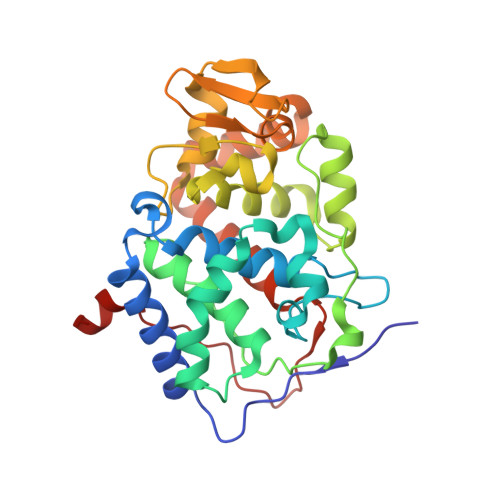
    Heme enzymes are ubiquitous in biology and catalyze a vast array of biological redox processes. The formation of high valent ferryl intermediates of the heme iron (known as Compounds I and Compound II) is implicated for a number of catalytic heme enzymes, but these species are formed only transiently and thus have proved somewhat elusive. In consequence, there has been conflicting evidence as to the nature of these ferryl intermediates in a number of different heme enzymes, in particular the precise nature of the bond between the heme iron and the bound oxygen atom. In this work, we present high resolution crystal structures of both Compound I and Compound II intermediates in two different heme peroxidase enzymes, cytochrome c peroxidase and ascorbate peroxidase, allowing direct and accurate comparison of the bonding interactions in the different intermediates. A consistent picture emerges across all structures, showing lengthening of the ferryl oxygen bond (and presumed protonation) on reduction of Compound I to Compound II. These data clarify long standing inconsistencies on the nature of the ferryl heme species in these intermediates.


  • Organizational Affiliation

    Department of Chemistry, Henry Wellcome Building, University of Leicester, University Road, Leicester LE1 7RH, United Kingdom.


Macromolecules
Find similar proteins by:  (by identity cutoff)  |  3D Structure
Entity ID: 1
MoleculeChains Sequence LengthOrganismDetailsImage
CYTOCHROME C PEROXIDASE, MITOCHONDRIAL294Saccharomyces cerevisiaeMutation(s): 0 
EC: 1.11.1.5
UniProt
Find proteins for P00431 (Saccharomyces cerevisiae (strain ATCC 204508 / S288c))
Explore P00431 
Go to UniProtKB:  P00431
Entity Groups  
Sequence Clusters30% Identity50% Identity70% Identity90% Identity95% Identity100% Identity
UniProt GroupP00431
Sequence Annotations
Expand
  • Reference Sequence
Experimental Data & Validation

Experimental Data

  • Method: X-RAY DIFFRACTION
  • Resolution: 1.69 Å
  • R-Value Free: 0.210 
  • R-Value Work: 0.175 
  • R-Value Observed: 0.177 
  • Space Group: P 21 21 21
Unit Cell:
Length ( Å )Angle ( ˚ )
a = 51.053α = 90
b = 75.193β = 90
c = 106.95γ = 90
Software Package:
Software NamePurpose
REFMACrefinement
MOSFLMdata reduction
SCALAdata scaling
CCP4phasing

Structure Validation

View Full Validation Report


Ligand Structure Quality Assessment 


Entry History 

Deposition Data

Revision History  (Full details and data files)

  • Version 1.0: 2010-07-14
    Type: Initial release
  • Version 1.1: 2011-05-08
    Changes: Version format compliance
  • Version 1.2: 2011-07-13
    Changes: Version format compliance
  • Version 1.3: 2018-02-07
    Changes: Database references, Source and taxonomy
  • Version 1.4: 2023-12-20
    Changes: Data collection, Database references, Derived calculations, Other, Refinement description