2ZBF

Calcium pump crystal structure with bound BeF3 and TG in the absence of calcium


Experimental Data Snapshot

  • Method: X-RAY DIFFRACTION
  • Resolution: 2.40 Å
  • R-Value Free: 0.257 
  • R-Value Work: 0.212 
  • R-Value Observed: 0.212 

wwPDB Validation   3D Report Full Report


This is version 1.2 of the entry. See complete history


Literature

How processing of aspartylphosphate is coupled to lumenal gating of the ion pathway in the calcium pump

Toyoshima, C.Norimatsu, Y.Iwasawa, S.Tsuda, T.Ogawa, H.

(2007) Proc Natl Acad Sci U S A 104: 19831-19836

  • DOI: https://doi.org/10.1073/pnas.0709978104
  • Primary Citation of Related Structures:  
    2ZBE, 2ZBF, 2ZBG

  • PubMed Abstract: 

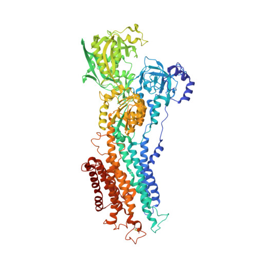
    Ca(2+)-ATPase of skeletal muscle sarcoplasmic reticulum is the best-studied member of the P-type or E1/E2 type ion transporting ATPases. It has been crystallized in seven different states that cover nearly the entire reaction cycle. Here we describe the structure of this ATPase complexed with phosphate analogs BeF(3)(-) and AlF(4)(-) in the absence of Ca(2+), which correspond to the E2P ground state and E2 approximately P transition state, respectively. The luminal gate is open with BeF(3)(-) and closed with AlF(4)(-). These and the E1 approximately P.ADP analog crystal structures show that a two-step rotation of the cytoplasmic A-domain opens and closes the luminal gate through the movements of the M1-M4 transmembrane helices. There are several conformational switches coupled to the rotation, and the one in the cytoplasmic part of M2 has critical importance. In the second step of rotation, positioning of one water molecule couples the hydrolysis of aspartylphosphate to closing of the gate.


  • Organizational Affiliation

    Institute of Molecular and Cellular Biosciences, University of Tokyo, 1-1-1 Yayoi, Bunkyo-ku, Tokyo 113-0032, Japan.


Macromolecules
Find similar proteins by:  (by identity cutoff)  |  3D Structure
Entity ID: 1
MoleculeChains Sequence LengthOrganismDetailsImage
Sarcoplasmic/endoplasmic reticulum calcium ATPase 1995Oryctolagus cuniculusMutation(s): 0 
EC: 3.6.3.8
Membrane Entity: Yes 
UniProt
Find proteins for P04191 (Oryctolagus cuniculus)
Explore P04191 
Go to UniProtKB:  P04191
Entity Groups  
Sequence Clusters30% Identity50% Identity70% Identity90% Identity95% Identity100% Identity
UniProt GroupP04191
Sequence Annotations
Expand
  • Reference Sequence
Small Molecules
Ligands 3 Unique
IDChains Name / Formula / InChI Key2D Diagram3D Interactions
TG1
Query on TG1

Download Ideal Coordinates CCD File 
D [auth A]OCTANOIC ACID [3S-[3ALPHA, 3ABETA, 4ALPHA, 6BETA, 6ABETA, 7BETA, 8ALPHA(Z), 9BALPHA]]-6-(ACETYLOXY)-2,3,-3A,4,5,6,6A,7,8,9B-DECAHYDRO-3,3A-DIHYDROXY-3,6,9-TRIMETHYL-8-[(2-METHYL-1-OXO-2-BUTENYL)OX Y]-2-OXO-4-(1-OXOBUTOXY)-AZULENO[4,5-B]FURAN-7-YL ESTER
C34 H50 O12
IXFPJGBNCFXKPI-FSIHEZPISA-N
BEF
Query on BEF

Download Ideal Coordinates CCD File 
C [auth A]BERYLLIUM TRIFLUORIDE ION
Be F3
OGIAHMCCNXDTIE-UHFFFAOYSA-K
MG
Query on MG

Download Ideal Coordinates CCD File 
B [auth A]MAGNESIUM ION
Mg
JLVVSXFLKOJNIY-UHFFFAOYSA-N
Binding Affinity Annotations 
IDSourceBinding Affinity
TG1 BindingDB:  2ZBF Ki: 0.1 (nM) from 1 assay(s)
Kd: 0.2 (nM) from 1 assay(s)
IC50: min: 0.2, max: 3.9 (nM) from 2 assay(s)
Experimental Data & Validation

Experimental Data

  • Method: X-RAY DIFFRACTION
  • Resolution: 2.40 Å
  • R-Value Free: 0.257 
  • R-Value Work: 0.212 
  • R-Value Observed: 0.212 
  • Space Group: P 21 21 2
Unit Cell:
Length ( Å )Angle ( ˚ )
a = 90.522α = 90
b = 136.473β = 90
c = 106.619γ = 90
Software Package:
Software NamePurpose
CNSrefinement
DENZOdata reduction
SCALEPACKdata scaling
CNSphasing

Structure Validation

View Full Validation Report


Entry History 

Deposition Data

Revision History  (Full details and data files)

  • Version 1.0: 2007-12-11
    Type: Initial release
  • Version 1.1: 2011-07-13
    Changes: Non-polymer description, Version format compliance
  • Version 1.2: 2023-11-01
    Changes: Data collection, Database references, Derived calculations, Refinement description, Structure summary