3CF2

Structure of P97/vcp in complex with ADP/AMP-PNP


Experimental Data Snapshot

  • Method: X-RAY DIFFRACTION
  • Resolution: 3.50 Å
  • R-Value Free: 0.285 
  • R-Value Work: 0.271 
  • R-Value Observed: 0.271 

wwPDB Validation   3D Report Full Report


Ligand Structure Quality Assessment 


This is version 1.3 of the entry. See complete history


Literature

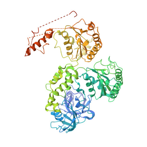
Improved structures of full-length p97, an AAA ATPase: implications for mechanisms of nucleotide-dependent conformational change.

Davies, J.M.Brunger, A.T.Weis, W.I.

(2008) Structure 16: 715-726

  • DOI: https://doi.org/10.1016/j.str.2008.02.010
  • Primary Citation of Related Structures:  
    3CF0, 3CF1, 3CF2, 3CF3

  • PubMed Abstract: 

    The ATPases associated with various cellular activities (AAA) protein p97 has been implicated in a variety of cellular processes, including endoplasmic reticulum-associated degradation and homotypic membrane fusion. p97 belongs to a subgroup of AAA proteins that contains two nucleotide binding domains, D1 and D2. We determined the crystal structure of D2 at 3.0 A resolution. This model enabled rerefinement of full-length p97 in different nucleotide states against previously reported low-resolution diffraction data to significantly improved R values and Ramachandran statistics. Although the overall fold remained similar, there are significant improvements, especially around the D2 nucleotide binding site. The rerefinement illustrates the importance of knowledge of high-resolution structures of fragments covering most of the whole molecule. The structures suggest that nucleotide hydrolysis is transformed into larger conformational changes by pushing of one D2 domain by its neighbor in the hexamer, and transmission of nucleotide-state information through the D1-D2 linker to displace the N-terminal, effector binding domain.


  • Organizational Affiliation

    Department of Structural Biology, Stanford University, Stanford, CA 94305-5432, USA.


Macromolecules
Find similar proteins by:  (by identity cutoff)  |  3D Structure
Entity ID: 1
MoleculeChains Sequence LengthOrganismDetailsImage
Transitional endoplasmic reticulum ATPase
A, B, C, D
806Mus musculusMutation(s): 0 
Gene Names: Vcp
UniProt
Find proteins for Q01853 (Mus musculus)
Explore Q01853 
Go to UniProtKB:  Q01853
Entity Groups  
Sequence Clusters30% Identity50% Identity70% Identity90% Identity95% Identity100% Identity
UniProt GroupQ01853
Sequence Annotations
Expand
  • Reference Sequence
Experimental Data & Validation

Experimental Data

  • Method: X-RAY DIFFRACTION
  • Resolution: 3.50 Å
  • R-Value Free: 0.285 
  • R-Value Work: 0.271 
  • R-Value Observed: 0.271 
  • Space Group: P 3
Unit Cell:
Length ( Å )Angle ( ˚ )
a = 144.9α = 90
b = 144.9β = 90
c = 164.4γ = 120
Software Package:
Software NamePurpose
CNSrefinement
MOSFLMdata reduction
SCALAdata scaling
CNSphasing

Structure Validation

View Full Validation Report


Ligand Structure Quality Assessment 


Entry History 

Deposition Data

Revision History  (Full details and data files)

  • Version 1.0: 2008-04-22
    Type: Initial release
  • Version 1.1: 2011-07-13
    Changes: Version format compliance
  • Version 1.2: 2012-03-07
    Changes: Other
  • Version 1.3: 2024-02-21
    Changes: Data collection, Database references, Derived calculations