3CWW

Crystal Structure of IDE-bradykinin complex


Experimental Data Snapshot

  • Method: X-RAY DIFFRACTION
  • Resolution: 1.96 Å
  • R-Value Free: 0.208 
  • R-Value Work: 0.181 
  • R-Value Observed: 0.183 

wwPDB Validation   3D Report Full Report


This is version 1.4 of the entry. See complete history


Literature

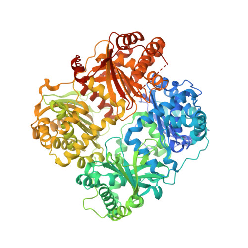
Molecular Bases for the Recognition of Short Peptide Substrates and Cysteine-Directed Modifications of Human Insulin-Degrading Enzyme

Malito, E.Ralat, L.A.Manolopoulou, M.Tsay, J.L.Wadlington, N.L.Tang, W.J.

(2008) Biochemistry 47: 12822-12834

  • DOI: https://doi.org/10.1021/bi801192h
  • Primary Citation of Related Structures:  
    3CWW

  • PubMed Abstract: 

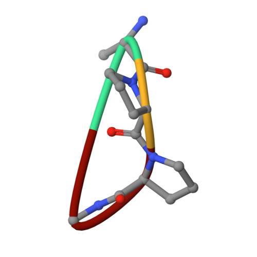
    Insulin degrading enzyme (IDE) utilizes a large catalytic chamber to selectively bind and degrade peptide substrates such as insulin and amyloid beta (Abeta). Tight interactions with substrates occur at an exosite located approximately 30 A away from the catalytic center that anchors the N-terminus of substrates to facilitate binding and subsequent cleavages at the catalytic site. However, IDE also degrades peptide substrates that are too short to occupy both the catalytic site and the exosite simultaneously. Here, we use kinins as a model system to address the kinetics and regulation of human IDE with short peptides. IDE specifically degrades bradykinin and kallidin at the Pro/Phe site. A 1.9 A crystal structure of bradykinin-bound IDE reveals the binding of bradykinin to the exosite and not to the catalytic site. In agreement with observed high K(m) values, this suggests low affinity of bradykinin for IDE. This structure also provides the molecular basis on how the binding of short peptides at the exosite could regulate substrate recognition. We also found that human IDE is potently inhibited by physiologically relevant concentrations of S-nitrosylation and oxidation agents. Cysteine-directed modifications play a key role, since an IDE mutant devoid of all 13 cysteines is insensitive to the inhibition by S-nitrosoglutathione, hydrogen peroxide, or N-ethylmaleimide. Specifically, cysteine 819 of human IDE is located inside the catalytic chamber pointing toward an extended hydrophobic pocket and is critical for the inactivation. Thiol-directed modification of this residue likely causes local structural perturbation to reduce substrate binding and catalysis.


  • Organizational Affiliation

    Ben-May Department for Cancer Research, Biological Science Collegiate Division, and Committee on Neurobiology, The University of Chicago, Chicago, Illinois 60637, USA.


Macromolecules
Find similar proteins by:  (by identity cutoff)  |  3D Structure
Entity ID: 1
MoleculeChains Sequence LengthOrganismDetailsImage
Insulin-degrading enzyme
A, B
990Homo sapiensMutation(s): 32 
Gene Names: IDERP11-366I13.1-001hCG_1810909
EC: 3.4.24.56
UniProt & NIH Common Fund Data Resources
Find proteins for P14735 (Homo sapiens)
Explore P14735 
Go to UniProtKB:  P14735
PHAROS:  P14735
GTEx:  ENSG00000119912 
Entity Groups  
Sequence Clusters30% Identity50% Identity70% Identity90% Identity95% Identity100% Identity
UniProt GroupP14735
Sequence Annotations
Expand
  • Reference Sequence

Find similar proteins by:  Sequence   |   3D Structure  

Entity ID: 2
MoleculeChains Sequence LengthOrganismDetailsImage
bradykinin N-terminal tetrapeptide analogueC [auth D],
D [auth E]
4N/AMutation(s): 0 
Sequence Annotations
Expand
  • Reference Sequence
Small Molecules
Ligands 3 Unique
IDChains Name / Formula / InChI Key2D Diagram3D Interactions
DIO
Query on DIO

Download Ideal Coordinates CCD File 
H [auth A],
I [auth A],
M [auth B],
N [auth B],
O [auth B]
1,4-DIETHYLENE DIOXIDE
C4 H8 O2
RYHBNJHYFVUHQT-UHFFFAOYSA-N
ZN
Query on ZN

Download Ideal Coordinates CCD File 
E [auth A],
J [auth B]
ZINC ION
Zn
PTFCDOFLOPIGGS-UHFFFAOYSA-N
ACT
Query on ACT

Download Ideal Coordinates CCD File 
F [auth A],
G [auth A],
K [auth B],
L [auth B]
ACETATE ION
C2 H3 O2
QTBSBXVTEAMEQO-UHFFFAOYSA-M
Experimental Data & Validation

Experimental Data

  • Method: X-RAY DIFFRACTION
  • Resolution: 1.96 Å
  • R-Value Free: 0.208 
  • R-Value Work: 0.181 
  • R-Value Observed: 0.183 
  • Space Group: P 65
Unit Cell:
Length ( Å )Angle ( ˚ )
a = 262.448α = 90
b = 262.448β = 90
c = 90.628γ = 120
Software Package:
Software NamePurpose
PHENIXrefinement
PDB_EXTRACTdata extraction
ADSCdata collection
DENZOdata reduction
SCALEPACKdata scaling
PHASERphasing

Structure Validation

View Full Validation Report


Entry History 

Deposition Data

Revision History  (Full details and data files)

  • Version 1.0: 2008-11-25
    Type: Initial release
  • Version 1.1: 2011-07-13
    Changes: Version format compliance
  • Version 1.2: 2017-10-25
    Changes: Refinement description
  • Version 1.3: 2021-10-20
    Changes: Database references, Derived calculations
  • Version 1.4: 2023-08-30
    Changes: Data collection, Refinement description