3D1F

Crystal structure of E. coli sliding clamp (beta) bound to a polymerase III peptide


Experimental Data Snapshot

  • Method: X-RAY DIFFRACTION
  • Resolution: 2.00 Å
  • R-Value Free: 0.255 
  • R-Value Work: 0.217 

wwPDB Validation   3D Report Full Report


Ligand Structure Quality Assessment 


This is version 1.4 of the entry. See complete history


Literature

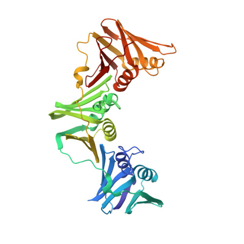
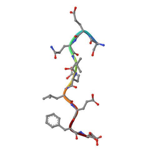
Structure of a small-molecule inhibitor of a DNA polymerase sliding clamp.

Georgescu, R.E.Yurieva, O.Kim, S.S.Kuriyan, J.Kong, X.P.O'Donnell, M.

(2008) Proc Natl Acad Sci U S A 105: 11116-11121

  • DOI: https://doi.org/10.1073/pnas.0804754105
  • Primary Citation of Related Structures:  
    3D1E, 3D1F, 3D1G

  • PubMed Abstract: 

    DNA polymerases attach to the DNA sliding clamp through a common overlapping binding site. We identify a small-molecule compound that binds the protein-binding site in the Escherichia coli beta-clamp and differentially affects the activity of DNA polymerases II, III, and IV. To understand the molecular basis of this discrimination, the cocrystal structure of the chemical inhibitor is solved in complex with beta and is compared with the structures of Pol II, Pol III, and Pol IV peptides bound to beta. The analysis reveals that the small molecule localizes in a region of the clamp to which the DNA polymerases attach in different ways. The results suggest that the small molecule may be useful in the future to probe polymerase function with beta, and that the beta-clamp may represent an antibiotic target.


  • Organizational Affiliation

    The Rockefeller University and Howard Hughes Medical Institute, 1230 York Avenue, P. O. Box 228, New York, NY 10065, USA.


Macromolecules
Find similar proteins by:  (by identity cutoff)  |  3D Structure
Entity ID: 1
MoleculeChains Sequence LengthOrganismDetailsImage
DNA polymerase III subunit beta
A, B
366Escherichia coliMutation(s): 0 
Gene Names: dnaN
EC: 2.7.7.7
UniProt
Find proteins for P0A988 (Escherichia coli (strain K12))
Explore P0A988 
Go to UniProtKB:  P0A988
Entity Groups  
Sequence Clusters30% Identity50% Identity70% Identity90% Identity95% Identity100% Identity
UniProt GroupP0A988
Sequence Annotations
Expand
  • Reference Sequence

Find similar proteins by:  Sequence   |   3D Structure  

Entity ID: 2
MoleculeChains Sequence LengthOrganismDetailsImage
Nonapeptide from polymerase III C-terminalC [auth P],
D [auth Q]
9N/AMutation(s): 0 
Sequence Annotations
Expand
  • Reference Sequence
Experimental Data & Validation

Experimental Data

  • Method: X-RAY DIFFRACTION
  • Resolution: 2.00 Å
  • R-Value Free: 0.255 
  • R-Value Work: 0.217 
  • Space Group: P 32
Unit Cell:
Length ( Å )Angle ( ˚ )
a = 65.778α = 90
b = 65.778β = 90
c = 209.472γ = 120
Software Package:
Software NamePurpose
DENZOdata reduction
SCALEPACKdata scaling
MOLREPphasing
CNSrefinement
PDB_EXTRACTdata extraction
ADSCdata collection

Structure Validation

View Full Validation Report


Ligand Structure Quality Assessment 


Entry History 

Deposition Data

Revision History  (Full details and data files)

  • Version 1.0: 2008-07-29
    Type: Initial release
  • Version 1.1: 2011-07-13
    Changes: Version format compliance
  • Version 1.2: 2017-10-25
    Changes: Refinement description
  • Version 1.3: 2018-01-24
    Changes: Structure summary
  • Version 1.4: 2023-08-30
    Changes: Data collection, Database references, Derived calculations, Refinement description