3D6G

Fc fragment of IgG1 (Herceptin) with protein-A mimetic peptide dendrimer ligand.


Experimental Data Snapshot

  • Method: X-RAY DIFFRACTION
  • Resolution: 2.30 Å
  • R-Value Free: 0.257 
  • R-Value Work: 0.188 
  • R-Value Observed: 0.190 

wwPDB Validation   3D Report Full Report


Ligand Structure Quality Assessment 


This is version 2.1 of the entry. See complete history


Literature

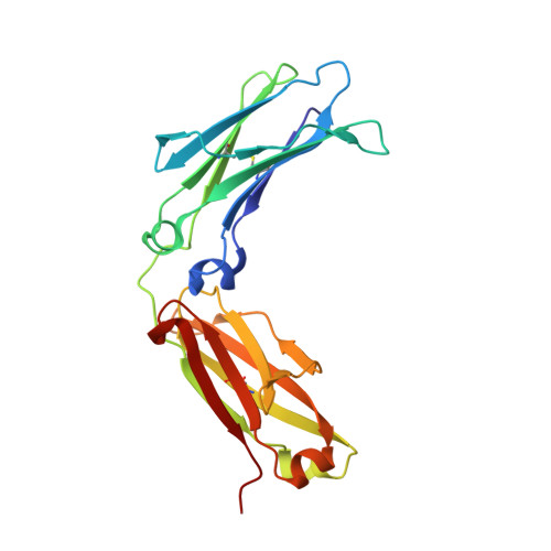
Structural characterization of a Protein A mimetic peptide dendrimer bound to human IgG.

Moiani, D.Salvalaglio, M.Cavallotti, C.Bujacz, A.Redzynia, I.Bujacz, G.Dinon, F.Pengo, P.Fassina, G.

(2009) J Phys Chem B 113: 16268-16275

  • DOI: https://doi.org/10.1021/jp909405b
  • Primary Citation of Related Structures:  
    3D6G

  • PubMed Abstract: 

    Understanding the chemical physical properties of protein binding sites is at the basis of the rational design of protein ligands. The hinge region of the Fc fragment of immunoglobulin G is an important and well characterized protein binding site, known to interact with several natural proteins and synthetic ligands. Here, we report structural evidence that a Staphylococcus aureus Protein A mimetic peptide dendrimer, deduced by a combinatorial approach, binds close to the Cgamma2/Cgamma3 interface of the constant fragment of a human IgG1 molecule, partially hindering the Protein A binding site. The X-ray analysis evidenced a primary binding site located between a terminal Arg residue of the ligand peptidic arm and a hydrophobic protein site consisting of Val308, Leu309, and His310. A molecular dynamic analysis of the model derived from the X-ray structure showed that in water at room temperature the complex is further stabilized by the formation of at least one more contact between a terminal Arg residue of the second arm of the peptide and the carboxylic group of a protein amino acid, such as Glu318, Asp312, or Asp280. It appears thus that stability of the Fc-dendrimer complex is determined by the synergetic formation of multiple bonds of different nature between the dendrimer arms and the protein accessible sites. The electrostatic and van der Waals energies of the complex were monitored during the MD simulations and confirmed the energetic stability of the two interactions.


  • Organizational Affiliation

    Department of Chimica, Materiali e Ingegneria Chimica, Politecnico di Milano, 20131 Milano, Italy.


Macromolecules
Find similar proteins by:  (by identity cutoff)  |  3D Structure
Entity ID: 1
MoleculeChains Sequence LengthOrganismDetailsImage
Ig gamma-1 chain C region
A, B
210Homo sapiensMutation(s): 0 
Gene Names: IGHG1
UniProt & NIH Common Fund Data Resources
Find proteins for P01857 (Homo sapiens)
Explore P01857 
Go to UniProtKB:  P01857
PHAROS:  P01857
GTEx:  ENSG00000211896 
Entity Groups  
Sequence Clusters30% Identity50% Identity70% Identity90% Identity95% Identity100% Identity
UniProt GroupP01857
Sequence Annotations
Expand
  • Reference Sequence
Oligosaccharides

Help

Entity ID: 2
MoleculeChains Length2D Diagram Glycosylation3D Interactions
2-acetamido-2-deoxy-beta-D-glucopyranose-(1-2)-alpha-D-mannopyranose-(1-3)-[2-acetamido-2-deoxy-beta-D-glucopyranose-(1-2)-alpha-D-mannopyranose-(1-6)]beta-D-mannopyranose-(1-4)-2-acetamido-2-deoxy-beta-D-glucopyranose-(1-4)-[alpha-L-fucopyranose-(1-6)]2-acetamido-2-deoxy-beta-D-glucopyranose
C, D
8N-Glycosylation
Glycosylation Resources
GlyTouCan:  G80858MF
GlyCosmos:  G80858MF
GlyGen:  G80858MF
Small Molecules
Ligands 1 Unique
IDChains Name / Formula / InChI Key2D Diagram3D Interactions
X12
Query on X12

Download Ideal Coordinates CCD File 
E [auth B]2-[[(2S)-2,6-bis[[(2S)-2,6-bis[[(2R)-2-[[(2R,3R)-2-[[(2R)-2-amino-5-carbamimidamido-pentanoyl]amino]-3-hydroxy-butanoyl]amino]-3-(4-hydroxyphenyl)propanoyl]amino]hexanoyl]amino]hexanoyl]amino]ethanoic acid
C96 H153 N31 O25
GFRBISAVUSZQEP-GABXEQJVSA-N
Experimental Data & Validation

Experimental Data

  • Method: X-RAY DIFFRACTION
  • Resolution: 2.30 Å
  • R-Value Free: 0.257 
  • R-Value Work: 0.188 
  • R-Value Observed: 0.190 
  • Space Group: P 21 21 21
Unit Cell:
Length ( Å )Angle ( ˚ )
a = 49.778α = 90
b = 79.155β = 90
c = 139.223γ = 90
Software Package:
Software NamePurpose
REFMACrefinement
HKL-2000data collection
DENZOdata reduction
SCALEPACKdata scaling
MOLREPphasing

Structure Validation

View Full Validation Report


Ligand Structure Quality Assessment 


Entry History 

Deposition Data

Revision History  (Full details and data files)

  • Version 1.0: 2009-06-16
    Type: Initial release
  • Version 1.1: 2011-07-13
    Changes: Advisory, Version format compliance
  • Version 1.2: 2019-12-25
    Changes: Data collection, Database references, Derived calculations
  • Version 2.0: 2020-07-29
    Type: Remediation
    Reason: Carbohydrate remediation
    Changes: Advisory, Atomic model, Data collection, Derived calculations, Structure summary
  • Version 2.1: 2023-11-01
    Changes: Data collection, Database references, Derived calculations, Refinement description, Structure summary