p38alpha MAP kinase C-terminal domain binding pocket characterized by crystallographic and computational analyses.
Perry, J.J., Harris, R.M., Moiani, D., Olson, A.J., Tainer, J.A.(2009) J Mol Biol 391: 1-11
- PubMed: 19501598
- DOI: https://doi.org/10.1016/j.jmb.2009.06.005
- Primary Citation of Related Structures:
3HVC - PubMed Abstract:
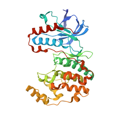
The mitogen-activated protein (MAP) kinase protein family has a critical role in cellular signaling events, with MAP kinase p38alpha acting in inflammatory processes and being an important drug discovery target. MAP kinase drug design efforts have focused on small-molecule inhibitors of the ATP catalytic site, which exhibit dose-limiting adverse effects. Therefore, characterizing other potential sites that bind substrates, inhibitors, or allosteric effectors is of great interest. Here, we present the crystal structure of human p38alpha MAP kinase, which has a lead compound bound both in the active site and in the lipid-binding site of the C-terminal cap. This C-terminal cap is formed from an extension to the kinase fold, unique to the MAP kinase and cyclin-dependent kinase families and glycogen synthase kinase 3. Binding of this lead, 4-[3-(4-fluorophenyl)-1H-pyrazol-4-yl]pyridine, to wild-type p38alpha induces movement of the C-terminal cap region, creating a hydrophobic pocket centered around residue Trp197. Computational analysis of this C-terminal domain pocket indicates notable flexibility for potentially binding different-shaped compounds, including lipids, oxidized arachidonic acid species such as leukotrienes, and small-molecule effectors. Furthermore, our structural results defining the open p38alpha C-lobe pocket provide a detailed framework for the design of novel small molecules with affinities comparable to active-site binders: to bind and potentially modulate the shape and activity of p38alpha in predetermined ways. Moreover, these results and analyses of p38alpha suggest strategies for designing specific binding compounds applicable to other MAP kinases, as well as the cyclin-dependent kinase family and glycogen synthase kinase 3beta that also utilize the C-terminal insert in their interactions.
Organizational Affiliation:
Department of Molecular Biology, The Scripps Research Institute, La Jolla, CA 92037, USA.