3T4Y

S25-2- KDO monosaccharide complex


Experimental Data Snapshot

  • Method: X-RAY DIFFRACTION
  • Resolution: 1.73 Å
  • R-Value Free: 0.225 
  • R-Value Work: 0.195 
  • R-Value Observed: 0.198 

wwPDB Validation   3D Report Full Report


Ligand Structure Quality Assessment 


This is version 1.2 of the entry. See complete history


Literature

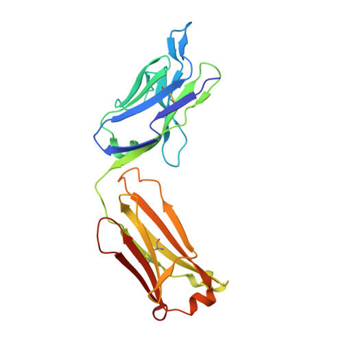
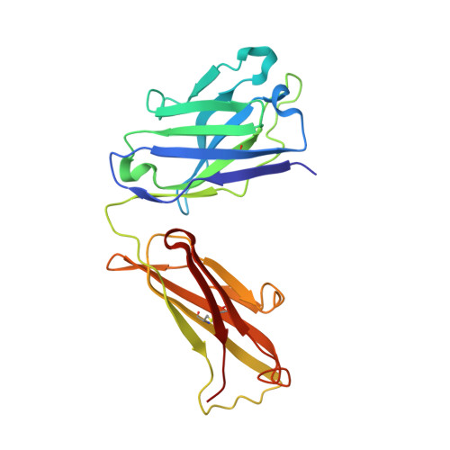
Germline antibody recognition of distinct carbohydrate epitopes.

Nguyen, H.P.Seto, N.O.MacKenzie, C.R.Brade, L.Kosma, P.Brade, H.Evans, S.V.

(2003) Nat Struct Biol 10: 1019-1025

  • DOI: https://doi.org/10.1038/nsb1014
  • Primary Citation of Related Structures:  
    1Q9K, 1Q9L, 1Q9O, 1Q9W, 3SY0, 3T4Y, 3T65, 3T77

  • PubMed Abstract: 

    High-resolution structures reveal how a germline antibody can recognize a range of clinically relevant carbohydrate epitopes. The germline response to a carbohydrate immunogen can be critical to survivability, with selection for antibody gene segments that both confer protection against common pathogens and retain the flexibility to adapt to new disease organisms. We show here that antibody S25-2 binds several distinct inner-core epitopes of bacterial lipopolysaccharides (LPSs) by linking an inherited monosaccharide residue binding site with a subset of complementarity-determining regions (CDRs) of limited flexibility positioned to recognize the remainder of an array of different epitopes. This strategy allows germline antibodies to adapt to different epitopes while minimizing entropic penalties associated with the immobilization of labile CDRs upon binding of antigen, and provides insight into the link between the genetic origin of individual CDRs and their respective roles in antigen recognition.


  • Organizational Affiliation

    Department of Biochemistry, Microbiology and Immunology, University of Ottawa, 451 Smyth Road, Ottawa, Ontario K1H 8M5, Canada.


Macromolecules
Find similar proteins by:  (by identity cutoff)  |  3D Structure
Entity ID: 1
MoleculeChains Sequence LengthOrganismDetailsImage
S25-2 FAB (IGG1K) LIGHT CHAIN219Mus musculusMutation(s): 0 
Entity Groups  
Sequence Clusters30% Identity50% Identity70% Identity90% Identity95% Identity100% Identity
Sequence Annotations
Expand
  • Reference Sequence
Find similar proteins by:  (by identity cutoff)  |  3D Structure
Entity ID: 2
MoleculeChains Sequence LengthOrganismDetailsImage
S25-2 FAB (IGG1K) HEAVY CHAIN222Mus musculusMutation(s): 0 
Entity Groups  
Sequence Clusters30% Identity50% Identity70% Identity90% Identity95% Identity100% Identity
Sequence Annotations
Expand
  • Reference Sequence
Experimental Data & Validation

Experimental Data

  • Method: X-RAY DIFFRACTION
  • Resolution: 1.73 Å
  • R-Value Free: 0.225 
  • R-Value Work: 0.195 
  • R-Value Observed: 0.198 
  • Space Group: P 21 21 21
Unit Cell:
Length ( Å )Angle ( ˚ )
a = 45.8α = 90
b = 81.4β = 90
c = 132.4γ = 90
Software Package:
Software NamePurpose
PHENIXrefinement
PDB_EXTRACTdata extraction

Structure Validation

View Full Validation Report


Ligand Structure Quality Assessment 


Entry History 

Deposition Data

Revision History  (Full details and data files)

  • Version 1.0: 2011-08-03
    Type: Initial release
  • Version 1.1: 2020-07-29
    Type: Remediation
    Reason: Carbohydrate remediation
    Changes: Data collection, Derived calculations, Structure summary
  • Version 1.2: 2023-09-13
    Changes: Data collection, Database references, Refinement description, Structure summary