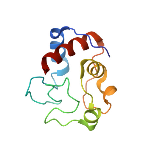
3TYI

Crystal Structure of Cytochrome c - p-Sulfonatocalix[4]arene Complexes


Experimental Data Snapshot

  • Method: X-RAY DIFFRACTION
  • Resolution: 1.40 Å
  • R-Value Free: 0.198 
  • R-Value Work: 0.177 
  • R-Value Observed: 0.177 

wwPDB Validation   3D Report Full Report


Ligand Structure Quality Assessment 


This is version 1.3 of the entry. See complete history


Literature

Protein camouflage in cytochrome c-calixarene complexes.

McGovern, R.E.Fernandes, H.Khan, A.R.Power, N.P.Crowley, P.B.

(2012) Nat Chem 4: 527-533

  • DOI: https://doi.org/10.1038/nchem.1342
  • Primary Citation of Related Structures:  
    3TYI

  • PubMed Abstract: 

    Small molecules that recognize protein surfaces are important tools for modifying protein interaction properties. Since the 1980s, several thousand studies concerning calixarenes and host-guest interactions have been published. Although there is growing interest in protein-calixarene interactions, only limited structural information has been available to date. We now report the crystal structure of a protein-calixarene complex. The water-soluble p-sulfonatocalix[4]arene is shown to bind the lysine-rich cytochrome c at three different sites. Binding curves obtained from NMR titrations reveal an interaction process that involves two or more binding sites. Together, the data indicate a dynamic complex in which the calixarene explores the surface of cytochrome c. In addition to providing valuable information on protein recognition, the data also indicate that the calixarene is a mediator of protein-protein interactions, with potential applications in generating assemblies and promoting crystallization.


  • Organizational Affiliation

    School of Chemistry, National University of Ireland Galway, University Road, Galway, Ireland.


Macromolecules
Find similar proteins by:  (by identity cutoff)  |  3D Structure
Entity ID: 1
MoleculeChains Sequence LengthOrganismDetailsImage
Cytochrome c iso-1
A, B
108Saccharomyces cerevisiae S288CMutation(s): 1 
Gene Names: CYC1YJR048WJ1653
UniProt
Find proteins for P00044 (Saccharomyces cerevisiae (strain ATCC 204508 / S288c))
Explore P00044 
Go to UniProtKB:  P00044
Entity Groups  
Sequence Clusters30% Identity50% Identity70% Identity90% Identity95% Identity100% Identity
UniProt GroupP00044
Sequence Annotations
Expand
  • Reference Sequence
Small Molecules
Ligands 2 Unique
IDChains Name / Formula / InChI Key2D Diagram3D Interactions
T3Y
Query on T3Y

Download Ideal Coordinates CCD File 
D [auth A],
F [auth B],
G [auth B]
25,26,27,28-tetrahydroxypentacyclo[19.3.1.1~3,7~.1~9,13~.1~15,19~]octacosa-1(25),3(28),4,6,9(27),10,12,15(26),16,18,21,23-dodecaene-5,11,17,23-tetrasulfonic acid
C28 H24 O16 S4
JFYBCAFLVNKHHG-UHFFFAOYSA-N
HEM
Query on HEM

Download Ideal Coordinates CCD File 
C [auth A],
E [auth B]
PROTOPORPHYRIN IX CONTAINING FE
C34 H32 Fe N4 O4
KABFMIBPWCXCRK-RGGAHWMASA-L
Experimental Data & Validation

Experimental Data

  • Method: X-RAY DIFFRACTION
  • Resolution: 1.40 Å
  • R-Value Free: 0.198 
  • R-Value Work: 0.177 
  • R-Value Observed: 0.177 
  • Space Group: P 21 21 21
Unit Cell:
Length ( Å )Angle ( ˚ )
a = 36.044α = 90
b = 55.317β = 90
c = 117.419γ = 90
Software Package:
Software NamePurpose
MxCuBEdata collection
PHASERphasing
REFMACrefinement
HKL-2000data reduction
SCALAdata scaling

Structure Validation

View Full Validation Report


Ligand Structure Quality Assessment 


Entry History 

Deposition Data

Revision History  (Full details and data files)

  • Version 1.0: 2012-05-02
    Type: Initial release
  • Version 1.1: 2012-08-08
    Changes: Database references
  • Version 1.2: 2018-01-24
    Changes: Structure summary
  • Version 1.3: 2023-09-13
    Changes: Data collection, Database references, Derived calculations, Refinement description