4AP0

The mitotic kinesin Eg5 in complex with Mg-ADP and ispinesib


Experimental Data Snapshot

  • Method: X-RAY DIFFRACTION
  • Resolution: 2.59 Å
  • R-Value Free: 0.253 
  • R-Value Work: 0.203 
  • R-Value Observed: 0.206 

wwPDB Validation   3D Report Full Report


Ligand Structure Quality Assessment 


This is version 1.3 of the entry. See complete history


Literature

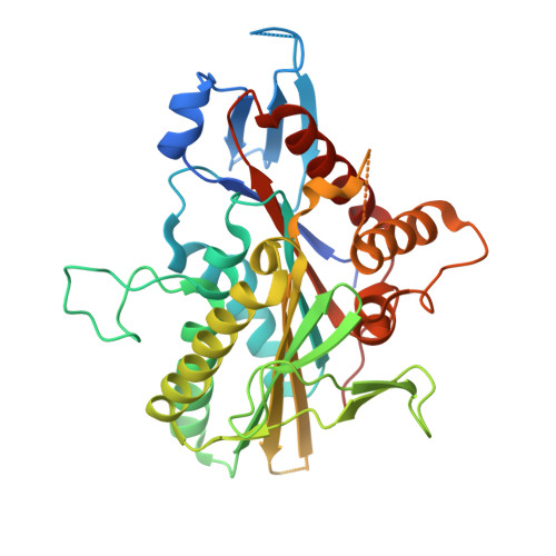
The Structure of the Ternary Eg5-Adp-Ispinesib Complex

Talapatra, S.K.Schuettelkopf, A.W.Kozielski, F.

(2012) Acta Crystallogr D Biol Crystallogr 68: 1311

  • DOI: https://doi.org/10.1107/S0907444912027965
  • Primary Citation of Related Structures:  
    4AP0

  • PubMed Abstract: 

    The human kinesin Eg5 is responsible for bipolar spindle formation during early mitosis. Inhibition of Eg5 triggers the formation of monoastral spindles, leading to mitotic arrest that eventually causes apoptosis. There is increasing evidence that Eg5 constitutes a potential drug target for the development of cancer chemotherapeutics. The most advanced Eg5-targeting agent is ispinesib, which exhibits potent antitumour activity and is currently in multiple phase II clinical trials. In this study, the crystal structure of the Eg5 motor domain in complex with ispinesib, supported by kinetic and thermodynamic binding data, is reported. Ispinesib occupies the same induced-fit pocket in Eg5 as other allosteric inhibitors, making extensive hydrophobic interactions with the protein. The data for the Eg5-ADP-ispinesib complex suffered from pseudo-merohedral twinning and revealed translational noncrystallographic symmetry, leading to challenges in data processing, space-group assignment and structure solution as well as in refinement. These complications may explain the lack of available structural information for this important agent and its analogues. The present structure represents the best interpretation of these data based on extensive data-reduction, structure-solution and refinement trials.


  • Organizational Affiliation

    Molecular Motor Laboratory, The Beatson Institute for Cancer Research, Garscube Estate, Switchback Road, Bearsden, Glasgow G61 1BD, Scotland, UK. s.talapatra@beatson.gla.ac.uk


Macromolecules
Find similar proteins by:  (by identity cutoff)  |  3D Structure
Entity ID: 1
MoleculeChains Sequence LengthOrganismDetailsImage
KINESIN-LIKE PROTEIN KIF11
A, B, C, D
370Homo sapiensMutation(s): 0 
UniProt & NIH Common Fund Data Resources
Find proteins for P52732 (Homo sapiens)
Explore P52732 
Go to UniProtKB:  P52732
PHAROS:  P52732
GTEx:  ENSG00000138160 
Entity Groups  
Sequence Clusters30% Identity50% Identity70% Identity90% Identity95% Identity100% Identity
UniProt GroupP52732
Sequence Annotations
Expand
  • Reference Sequence
Small Molecules
Ligands 4 Unique
IDChains Name / Formula / InChI Key2D Diagram3D Interactions
G7X
Query on G7X

Download Ideal Coordinates CCD File 
I [auth A],
O [auth B],
R [auth C],
V [auth D]
ISPINESIB MESILATE
C30 H33 Cl N4 O2
QJZRFPJCWMNVAV-HHHXNRCGSA-N
ADP
Query on ADP

Download Ideal Coordinates CCD File 
F [auth A],
K [auth B],
Q [auth C],
T [auth D]
ADENOSINE-5'-DIPHOSPHATE
C10 H15 N5 O10 P2
XTWYTFMLZFPYCI-KQYNXXCUSA-N
CL
Query on CL

Download Ideal Coordinates CCD File 
G [auth A]
H [auth A]
L [auth B]
M [auth B]
N [auth B]
G [auth A],
H [auth A],
L [auth B],
M [auth B],
N [auth B],
U [auth D]
CHLORIDE ION
Cl
VEXZGXHMUGYJMC-UHFFFAOYSA-M
MG
Query on MG

Download Ideal Coordinates CCD File 
E [auth A],
J [auth B],
P [auth C],
S [auth D]
MAGNESIUM ION
Mg
JLVVSXFLKOJNIY-UHFFFAOYSA-N
Binding Affinity Annotations 
IDSourceBinding Affinity
G7X BindingDB:  4AP0 Ki: min: 2.3, max: 2.5 (nM) from 2 assay(s)
IC50: min: 1.2, max: 47 (nM) from 3 assay(s)
PDBBind:  4AP0 Kd: 10 (nM) from 1 assay(s)
Experimental Data & Validation

Experimental Data

  • Method: X-RAY DIFFRACTION
  • Resolution: 2.59 Å
  • R-Value Free: 0.253 
  • R-Value Work: 0.203 
  • R-Value Observed: 0.206 
  • Space Group: P 1 21 1
Unit Cell:
Length ( Å )Angle ( ˚ )
a = 64.723α = 90
b = 112.607β = 90.02
c = 106.913γ = 90
Software Package:
Software NamePurpose
PHENIXrefinement
XDSdata reduction
XSCALEdata scaling
MOLREPphasing

Structure Validation

View Full Validation Report


Ligand Structure Quality Assessment 


Entry History 

Deposition Data

Revision History  (Full details and data files)

  • Version 1.0: 2012-09-26
    Type: Initial release
  • Version 1.1: 2012-10-03
    Changes: Database references
  • Version 1.2: 2019-05-08
    Changes: Data collection, Experimental preparation, Other
  • Version 1.3: 2023-12-20
    Changes: Data collection, Database references, Derived calculations, Other, Refinement description