4HGW

Crystal structure of S25-2 in complex with a 5,6-dehydro-Kdo disaccharide


Experimental Data Snapshot

  • Method: X-RAY DIFFRACTION
  • Resolution: 1.65 Å
  • R-Value Free: 0.198 
  • R-Value Work: 0.176 
  • R-Value Observed: 0.177 

wwPDB Validation   3D Report Full Report


This is version 2.1 of the entry. See complete history


Literature

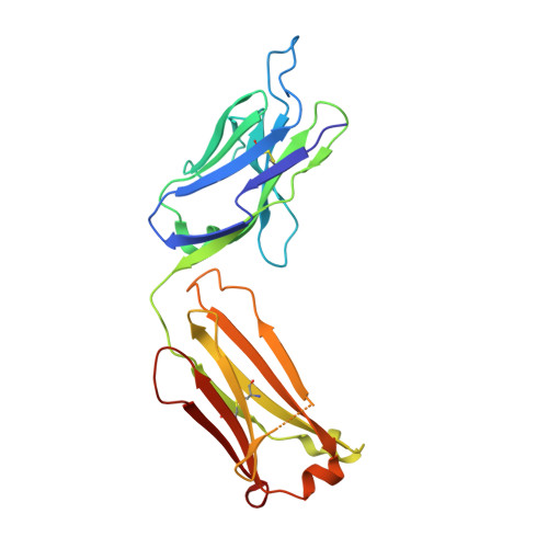
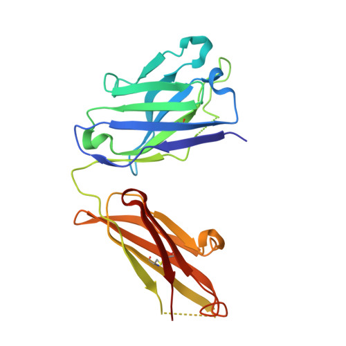
Exploring the cross-reactivity of S25-2: complex with a 5,6-dehydro-Kdo disaccharide.

Brooks, C.L.Wimmer, K.Kosma, P.Muller-Loennies, S.Brade, L.Brade, H.Evans, S.V.

(2013) Acta Crystallogr Sect F Struct Biol Cryst Commun 69: 2-5

  • DOI: https://doi.org/10.1107/S1744309112047422
  • Primary Citation of Related Structures:  
    4HGW

  • PubMed Abstract: 

    The near-germline antibody S25-2 exhibits a remarkable cross-reactivity for oligosaccharides containing the bacterial lipopolysaccharide carbohydrate 3-deoxy-D-manno-oct-2-ulosonic acid (Kdo). The recent synthesis of a variety of Kdo analogues permits a detailed structural analysis of the importance of specific interactions in antigen recognition by S25-2. The Kdo disaccharide analogue Kdo-(2→4)-5,6-dehydro-Kdo lacks a 5-OH group on the second Kdo residue and has been cocrystallized with S25-2. The structure reveals that the modification of the Kdo residue at position 5 results in a rearrangement of intramolecular hydrogen bonds in the antigen that allows it to assume a novel conformation in the antibody-combining site. The cross-reactive binding of S25-2 to this synthetic ligand highlights the adaptability of this antibody to non-natural synthetic analogues.


  • Organizational Affiliation

    Department of Biochemistry and Microbiology, University of Victoria, PO Box 3055 STN CSC, Victoria, BC V8W 3P6, Canada. cbrooks1@ualberta.ca


Macromolecules
Find similar proteins by:  (by identity cutoff)  |  3D Structure
Entity ID: 1
MoleculeChains Sequence LengthOrganismDetailsImage
Antibody Fab fragment, light chain219Mus musculusMutation(s): 0 
Entity Groups  
Sequence Clusters30% Identity50% Identity70% Identity90% Identity95% Identity100% Identity
Sequence Annotations
Expand
  • Reference Sequence
Find similar proteins by:  (by identity cutoff)  |  3D Structure
Entity ID: 2
MoleculeChains Sequence LengthOrganismDetailsImage
Antibody Fab fragment, heavy chain222Mus musculusMutation(s): 0 
Entity Groups  
Sequence Clusters30% Identity50% Identity70% Identity90% Identity95% Identity100% Identity
Sequence Annotations
Expand
  • Reference Sequence
Oligosaccharides

Help

Entity ID: 3
MoleculeChains Length2D Diagram Glycosylation3D Interactions
3-deoxy-alpha-D-manno-oct-2-ulopyranosonic acid-(2-4)-prop-2-en-1-yl 3,5-dideoxy-alpha-D-threo-oct-5-en-2-ulopyranosidonic acid
C
2N/AN/A
Glycosylation Resources
GlyTouCan:  G87312LN
GlyCosmos:  G87312LN
Experimental Data & Validation

Experimental Data

  • Method: X-RAY DIFFRACTION
  • Resolution: 1.65 Å
  • R-Value Free: 0.198 
  • R-Value Work: 0.176 
  • R-Value Observed: 0.177 
  • Space Group: P 21 21 21
Unit Cell:
Length ( Å )Angle ( ˚ )
a = 45.71α = 90
b = 81.3β = 90
c = 131.42γ = 90
Software Package:
Software NamePurpose
d*TREKdata reduction
PHENIXrefinement
PDB_EXTRACTdata extraction
d*TREKdata scaling
PHASERphasing

Structure Validation

View Full Validation Report


Entry History 

Deposition Data

Revision History  (Full details and data files)

  • Version 1.0: 2013-01-09
    Type: Initial release
  • Version 1.1: 2013-01-30
    Changes: Database references
  • Version 1.2: 2020-04-22
    Changes: Data collection, Derived calculations
  • Version 2.0: 2020-07-29
    Type: Remediation
    Reason: Carbohydrate remediation
    Changes: Atomic model, Data collection, Derived calculations, Structure summary
  • Version 2.1: 2023-09-20
    Changes: Data collection, Database references, Refinement description, Structure summary