4HZL

Neutralizing antibody mAb#8 in complex with the Epitope II of HCV E2 envelope protein


Experimental Data Snapshot

  • Method: X-RAY DIFFRACTION
  • Resolution: 2.85 Å
  • R-Value Free: 0.277 
  • R-Value Work: 0.221 
  • R-Value Observed: 0.224 

wwPDB Validation   3D Report Full Report


This is version 1.2 of the entry. See complete history


Literature

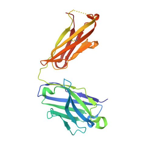
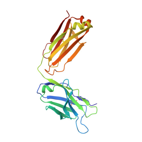
Structural evidence for a bifurcated mode of action in the antibody-mediated neutralization of hepatitis C virus.

Deng, L.Zhong, L.Struble, E.Duan, H.Ma, L.Harman, C.Yan, H.Virata-Theimer, M.L.Zhao, Z.Feinstone, S.Alter, H.Zhang, P.

(2013) Proc Natl Acad Sci U S A 110: 7418-7422

  • DOI: https://doi.org/10.1073/pnas.1305306110
  • Primary Citation of Related Structures:  
    4HZL

  • PubMed Abstract: 

    Hepatitis C virus (HCV) envelope glycoprotein E2 has been considered as a major target for vaccine design. Epitope II, mapped between residues 427-446 within the E2 protein, elicits antibodies that are either neutralizing or nonneutralizing. The fundamental mechanism of antibody-mediated neutralization at epitope II remains to be defined at the atomic level. Here we report the crystal structure of the epitope II peptide in complex with a monoclonal antibody (mAb#8) capable of neutralizing HCV. The complex structure revealed that this neutralizing antibody engages epitope II via interactions with both the C-terminal α-helix and the N-terminal loop using a bifurcated mode of action. Our structural insights into the key determinants for the antibody-mediated neutralization may contribute to the immune prophylaxis of HCV infection and the development of an effective HCV vaccine.


  • Organizational Affiliation

    Division of Hematology, Office of Blood Research and Review, Center for Biologics Evaluation and Research, Food and Drug Administration, Bethesda, MD 20892, USA.


Macromolecules
Find similar proteins by:  (by identity cutoff)  |  3D Structure
Entity ID: 1
MoleculeChains Sequence LengthOrganismDetailsImage
Fab heavy chainA [auth H],
D [auth A]
222Mus musculusMutation(s): 0 
Entity Groups  
Sequence Clusters30% Identity50% Identity70% Identity90% Identity95% Identity100% Identity
Sequence Annotations
Expand
  • Reference Sequence
Find similar proteins by:  (by identity cutoff)  |  3D Structure
Entity ID: 2
MoleculeChains Sequence LengthOrganismDetailsImage
Fab light chainB [auth L],
E [auth B]
217Mus musculusMutation(s): 0 
Entity Groups  
Sequence Clusters30% Identity50% Identity70% Identity90% Identity95% Identity100% Identity
Sequence Annotations
Expand
  • Reference Sequence

Find similar proteins by:  Sequence   |   3D Structure  

Entity ID: 3
MoleculeChains Sequence LengthOrganismDetailsImage
E2 envelop proteinC [auth E],
F
17Recombinant Hepatitis C virus H77(5'UTR-NS2)/JFH1_V787A,Q1247LMutation(s): 0 
UniProt
Find proteins for F5BWY6 (Recombinant Hepatitis C virus H77(5'UTR-NS2)/JFH1_V787A,Q1247L)
Explore F5BWY6 
Go to UniProtKB:  F5BWY6
Entity Groups  
Sequence Clusters30% Identity50% Identity70% Identity90% Identity95% Identity100% Identity
UniProt GroupF5BWY6
Sequence Annotations
Expand
  • Reference Sequence
Experimental Data & Validation

Experimental Data

  • Method: X-RAY DIFFRACTION
  • Resolution: 2.85 Å
  • R-Value Free: 0.277 
  • R-Value Work: 0.221 
  • R-Value Observed: 0.224 
  • Space Group: P 43 21 2
Unit Cell:
Length ( Å )Angle ( ˚ )
a = 137.252α = 90
b = 137.252β = 90
c = 140.49γ = 90
Software Package:
Software NamePurpose
ADSCdata collection
PHASERphasing
REFMACrefinement
HKL-2000data reduction
HKL-2000data scaling

Structure Validation

View Full Validation Report


Entry History 

Deposition Data

Revision History  (Full details and data files)

  • Version 1.0: 2013-05-01
    Type: Initial release
  • Version 1.1: 2013-05-15
    Changes: Database references
  • Version 1.2: 2023-09-20
    Changes: Data collection, Database references, Derived calculations, Refinement description