4OMW

Crystal structure of goat beta-lactoglobulin (orthorhombic form)


Experimental Data Snapshot

  • Method: X-RAY DIFFRACTION
  • Resolution: 2.30 Å
  • R-Value Free: 0.267 
  • R-Value Work: 0.225 
  • R-Value Observed: 0.227 

wwPDB Validation   3D Report Full Report


Ligand Structure Quality Assessment 


This is version 1.4 of the entry. See complete history


Literature

Conformational variability of goat beta-lactoglobulin: Crystallographic and thermodynamic studies.

Loch, J.I.Bonarek, P.Polit, A.Swiatek, S.Czub, M.Ludwikowska, M.Lewinski, K.

(2014) Int J Biol Macromol 72C: 1283-1291

  • DOI: https://doi.org/10.1016/j.ijbiomac.2014.10.031
  • Primary Citation of Related Structures:  
    4OMW, 4OMX

  • PubMed Abstract: 

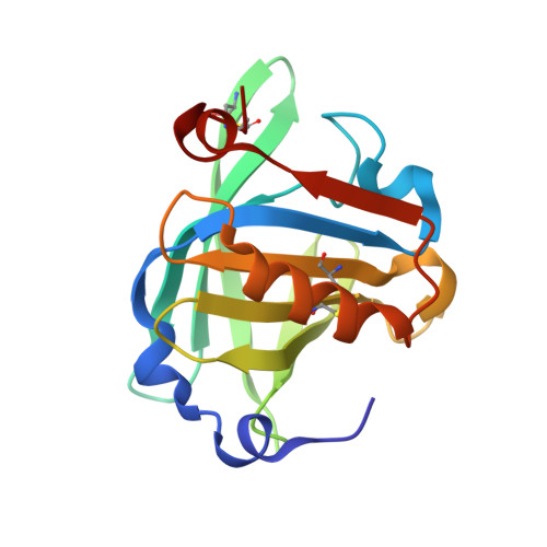
    Goat β-lactoglobulin (GLG), lipocalin protein sharing high sequence similarity to bovine β-lactoglobulin (BLG), has been structurally and thermodynamically characterized. Two crystal forms of GLG have been obtained, trigonal (P3121) and orthorhombic (P21212), with unique molecular packing, not observed previously for BLG. In the trigonal structure, GLG molecules have EF-loop in closed conformation while in the orthorhombic structure, for the first time, symmetric and asymmetric dimers of β-lactoglobulin are observed simultaneously. It indicates that the opening or closing EF-loop does not occur in both subunits at the same time but might be sequential and cooperative. Comparison of GLG and BLG structures revealed presence of various conformers of EF and GH. ITC studies showed that at pH 7.5 GLG binds sodium dodecyl sulfate with Gibbs energy similar to BLG, however, with different contribution from enthalpic and entropic component. At pH 7.5 GLG forms dimers with dimerization constant Ka = 34.28 × 10(3) M(-1), significantly higher than observed for BLG. Similar mechanism of conformational changes and ligand binding indicates that GLG and BLG may play analogous biological role.


  • Organizational Affiliation

    Department of Crystal Chemistry and Crystal Physics, Faculty of Chemistry, Jagiellonian University in Kraków, Ingardena 3, 30-060 Kraków, Poland.


Macromolecules
Find similar proteins by:  (by identity cutoff)  |  3D Structure
Entity ID: 1
MoleculeChains Sequence LengthOrganismDetailsImage
Beta-lactoglobulin
A, B, C, D
162Capra hircusMutation(s): 0 
UniProt
Find proteins for P02756 (Capra hircus)
Explore P02756 
Go to UniProtKB:  P02756
Entity Groups  
Sequence Clusters30% Identity50% Identity70% Identity90% Identity95% Identity100% Identity
UniProt GroupP02756
Sequence Annotations
Expand
  • Reference Sequence
Experimental Data & Validation

Experimental Data

  • Method: X-RAY DIFFRACTION
  • Resolution: 2.30 Å
  • R-Value Free: 0.267 
  • R-Value Work: 0.225 
  • R-Value Observed: 0.227 
  • Space Group: P 21 21 2
Unit Cell:
Length ( Å )Angle ( ˚ )
a = 150.05α = 90
b = 95.27β = 90
c = 55.35γ = 90
Software Package:
Software NamePurpose
MAR345data collection
PHASERphasing
REFMACrefinement
XDSdata reduction
SCALAdata scaling

Structure Validation

View Full Validation Report


Ligand Structure Quality Assessment 


Entry History 

Deposition Data

Revision History  (Full details and data files)

  • Version 1.0: 2014-11-19
    Type: Initial release
  • Version 1.1: 2014-12-17
    Changes: Database references
  • Version 1.2: 2015-02-18
    Changes: Non-polymer description
  • Version 1.3: 2017-11-22
    Changes: Data collection, Refinement description
  • Version 1.4: 2023-09-20
    Changes: Data collection, Database references, Derived calculations, Refinement description