4OT4

X-ray Structure of the Adduct formed between cisplatin and Ribonuclease A


Experimental Data Snapshot

  • Method: X-RAY DIFFRACTION
  • Resolution: 1.85 Å
  • R-Value Free: 0.247 
  • R-Value Work: 0.189 
  • R-Value Observed: 0.192 

wwPDB Validation   3D Report Full Report


This is version 1.2 of the entry. See complete history


Literature

Cisplatin binding to proteins: molecular structure of the ribonuclease a adduct.

Messori, L.Merlino, A.

(2014) Inorg Chem 53: 3929-3931

  • DOI: https://doi.org/10.1021/ic500360f
  • Primary Citation of Related Structures:  
    4OT4

  • PubMed Abstract: 

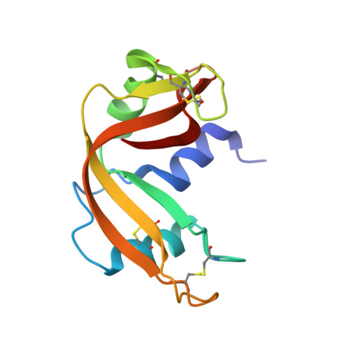
    The crystal structure of the main adduct formed in the reaction between cisplatin and bovine pancreatic ribonuclease is reported here. Notably, in both of the protein molecules present in the asymmetric unit, platinum(II) binding takes place exclusively at the level of Met29. In one of the two molecules, the Gln28 side chain completes the platinum coordination sphere, anchoring the cisplatin fragment to the protein in a bidentate fashion. These results contain interesting implications for understanding the biological chemistry of this important drug.


  • Organizational Affiliation

    Department of Chemistry, University of Florence , Via della Lastruccia 3, 50019 Sesto Fiorentino, Italy.


Macromolecules
Find similar proteins by:  (by identity cutoff)  |  3D Structure
Entity ID: 1
MoleculeChains Sequence LengthOrganismDetailsImage
Ribonuclease pancreatic
A, B
124Bos taurusMutation(s): 0 
Gene Names: RNASE1RNS1
EC: 3.1.27.5
UniProt
Find proteins for P61823 (Bos taurus)
Explore P61823 
Go to UniProtKB:  P61823
Entity Groups  
Sequence Clusters30% Identity50% Identity70% Identity90% Identity95% Identity100% Identity
UniProt GroupP61823
Sequence Annotations
Expand
  • Reference Sequence
Experimental Data & Validation

Experimental Data

  • Method: X-RAY DIFFRACTION
  • Resolution: 1.85 Å
  • R-Value Free: 0.247 
  • R-Value Work: 0.189 
  • R-Value Observed: 0.192 
  • Space Group: C 1 2 1
Unit Cell:
Length ( Å )Angle ( ˚ )
a = 99.767α = 90
b = 32.463β = 93.07
c = 67.575γ = 90
Software Package:
Software NamePurpose
CrystalCleardata collection
PHASERphasing
REFMACrefinement
HKL-2000data reduction
HKL-2000data scaling

Structure Validation

View Full Validation Report


Entry History 

Deposition Data

Revision History  (Full details and data files)

  • Version 1.0: 2014-07-09
    Type: Initial release
  • Version 1.1: 2014-07-16
    Changes: Derived calculations
  • Version 1.2: 2023-09-20
    Changes: Data collection, Database references, Derived calculations, Refinement description