4PQC

A sperm whale myoglobin single mutant F43H Mb with native His93 coordination


Experimental Data Snapshot

  • Method: X-RAY DIFFRACTION
  • Resolution: 1.50 Å
  • R-Value Free: 0.209 
  • R-Value Work: 0.181 
  • R-Value Observed: 0.183 

wwPDB Validation   3D Report Full Report


Ligand Structure Quality Assessment 


This is version 1.3 of the entry. See complete history


Literature

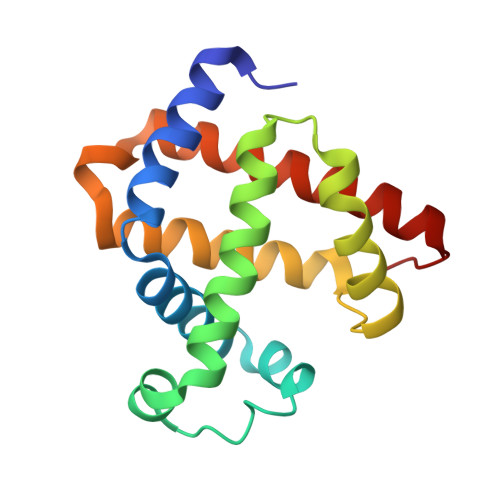
Regulating the coordination state of a heme protein by a designed distal hydrogen-bonding network.

Du, J.F.Li, W.Li, L.Wen, G.B.Lin, Y.W.Tan, X.

(2015) ChemistryOpen 4: 97-101

  • DOI: https://doi.org/10.1002/open.201402108
  • Primary Citation of Related Structures:  
    4PQ6, 4PQB, 4PQC

  • PubMed Abstract: 

    Heme coordination state determines the functional diversity of heme proteins. Using myoglobin as a model protein, we designed a distal hydrogen-bonding network by introducing both distal glutamic acid (Glu29) and histidine (His43) residues and regulated the heme into a bis-His coordination state with native ligands His64 and His93. This resembles the heme site in natural bis-His coordinated heme proteins such as cytoglobin and neuroglobin. A single mutation of L29E or F43H was found to form a distinct hydrogen-bonding network involving distal water molecules, instead of the bis-His heme coordination, which highlights the importance of the combination of multiple hydrogen-bonding interactions to regulate the heme coordination state. Kinetic studies further revealed that direct coordination of distal His64 to the heme iron negatively regulates fluoride binding and hydrogen peroxide activation by competing with the exogenous ligands. The new approach developed in this study can be generally applicable for fine-tuning the structure and function of heme proteins.


  • Organizational Affiliation

    School of Chemistry and Chemical Engineering, University of South China Hengyang, 421001, (P. R. China)).


Macromolecules
Find similar proteins by:  (by identity cutoff)  |  3D Structure
Entity ID: 1
MoleculeChains Sequence LengthOrganismDetailsImage
Myoglobin154Physeter catodonMutation(s): 1 
Gene Names: MB
UniProt
Find proteins for P02185 (Physeter macrocephalus)
Explore P02185 
Go to UniProtKB:  P02185
Entity Groups  
Sequence Clusters30% Identity50% Identity70% Identity90% Identity95% Identity100% Identity
UniProt GroupP02185
Sequence Annotations
Expand
  • Reference Sequence
Small Molecules
Ligands 1 Unique
IDChains Name / Formula / InChI Key2D Diagram3D Interactions
HEM
Query on HEM

Download Ideal Coordinates CCD File 
B [auth A]PROTOPORPHYRIN IX CONTAINING FE
C34 H32 Fe N4 O4
KABFMIBPWCXCRK-RGGAHWMASA-L
Experimental Data & Validation

Experimental Data

  • Method: X-RAY DIFFRACTION
  • Resolution: 1.50 Å
  • R-Value Free: 0.209 
  • R-Value Work: 0.181 
  • R-Value Observed: 0.183 
  • Space Group: P 21 21 21
Unit Cell:
Length ( Å )Angle ( ˚ )
a = 39.808α = 90
b = 48.055β = 90
c = 77.21γ = 90
Software Package:
Software NamePurpose
HKL-2000data collection
PHASERphasing
PHENIXrefinement
HKL-2000data reduction
HKL-2000data scaling

Structure Validation

View Full Validation Report


Ligand Structure Quality Assessment 


Entry History 

Deposition Data

Revision History  (Full details and data files)

  • Version 1.0: 2015-01-14
    Type: Initial release
  • Version 1.1: 2015-05-27
    Changes: Database references
  • Version 1.2: 2017-11-22
    Changes: Refinement description
  • Version 1.3: 2024-02-28
    Changes: Data collection, Database references, Derived calculations