Structure-activity correlations for three pyrido[2,3-d]pyrimidine antifolates binding to human and Pneumocystis carinii dihydrofolate reductase.
Cody, V., Pace, J., Namjoshi, O.A., Gangjee, A.(2015) Acta Crystallogr Sect F Struct Biol Cryst Commun 71: 799-803
- PubMed: 26057816
- DOI: https://doi.org/10.1107/S2053230X15008468
- Primary Citation of Related Structures:
4QHV, 4QJC, 4QJZ - PubMed Abstract:
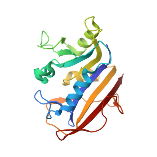
To further define the interactions that enhance the selectivity of binding and to directly compare the binding of the most potent analogue {N(6)-methyl-N(6)-(3,4,5-trifluorophenyl)pyrido[2,3-d]pyrimidine-2,4,6-triamine; compound 26} in the series of bicyclic pyrido[2,3-d]pyrimidine analogues of piritrexim (PTX) with native human (h), Pneumocystis carinii (pc) and Pneumocystis jirovecii (pj) dihydrofolate reductase (DHFR) enzymes, the crystal structures of hDHFR complexed with N(6)-methyl-N(6)-(4-isopropylphenyl)pyrido[2,3-d]pyrimidine-2,4,6-triamine (compound 22), of hDHFR complexed with compound 26 and of pcDHFR complexed with N(6)-methyl-N(6)-1-naphthylpyrido[2,3-d]pyrimidine-2,4,6-triamine (compound 24) are reported as ternary complexes with NADPH. This series of bicyclic pyrido[2,3-d]pyrimidines were designed in which there was a transposition of the 5-methyl group of PTX to the N9 position of the pyrido[2,3-d]pyrimidine. It was hypothesized that the N9-methyl group would preferentially interact with Ile123 of pcDHFR (and Ile123 of pjDHFR), but not with the shorter Val115 in hDHFR. Structure-activity data for this series of antifolates revealed that a trifluoro derivative (26) was the most selective against pjDHFR compared with mammalian DHFR (h/pj = 35.7). Structural data for the hDHFR-26 complex revealed that 26 binds in a different conformation from that observed in the pcDHFR-26 complex. In the hDHFR-26 complex the trifluorophenyl ring of 26 occupies a position near the cofactor-binding site, with close intermolecular contacts with Asp21, Ser59 and Ile60, whereas this ring in the pcDHFR-26 complex is positioned away from the cofactor site and near Ile65, with weaker contacts with Ile65, Phe69 and Ile123. Comparison of the intermolecular contacts between the N9-methyl group with Val115/Ile123 validates the hypothesis that the N9-methyl substituent preferentially interacts with Ile123 compared with Val115 of hDHFR, as the weaker contact with Val115 in the hDHFR structure is consistent with its weaker binding affinity compared with pcDHFR. The results for the structures of hDHFR-22 and pcDHFR-24 show that their inhibitor-binding orientation is similar to that observed in pcDHFR-26 and the pcDHFR variant (F69N) reported previously. The naphthyl moiety of 24 makes several intermolecular contacts with the active-site residues in pcDHFR that help to stabilize the binding, resulting in a more potent inhibitor.
Organizational Affiliation:
Structural Biology Department, Hauptman-Woodward Medical Research Institute, 700 Ellicott Street, Buffalo, NY 14203, USA.