4S18

The X-ray structure of the adduct formed in the reaction between bovine pancreatic ribonuclease and oxaliplatin


Experimental Data Snapshot

  • Method: X-RAY DIFFRACTION
  • Resolution: 2.27 Å
  • R-Value Free: 0.301 
  • R-Value Work: 0.225 
  • R-Value Observed: 0.228 

wwPDB Validation   3D Report Full Report


Ligand Structure Quality Assessment 


This is version 1.3 of the entry. See complete history


Literature

Interactions of carboplatin and oxaliplatin with proteins: Insights from X-ray structures and mass spectrometry studies of their ribonuclease A adducts.

Messori, L.Marzo, T.Merlino, A.

(2015) J Inorg Biochem 153: 136-142

  • DOI: https://doi.org/10.1016/j.jinorgbio.2015.07.011
  • Primary Citation of Related Structures:  
    4S0Q, 4S18

  • PubMed Abstract: 

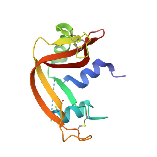
    Oxaliplatin and carboplatin are two platinum(II) drugs in widespread clinical use for the treatment of various types of cancers; yet, structural information on their interactions with proteins is scarce. Here, the X-ray structures of the adducts formed upon reaction of carboplatin and oxaliplatin with bovine pancreatic ribonuclease (RNase A) are reported and compared with results obtained for the structure of the RNase A-cisplatin adduct derived from isomorphous crystals, under the same experimental conditions. Additional details on the binding mode of these metallodrugs toward RNase A are provided by electrospray ionization mass spectrometry (ESI MS) measurements, thus offering insight on the occurring metal-protein interactions. Notably, while carboplatin and cisplatin mainly bind the side chain of Met29, oxaliplatin also binds the side chains of Asp14, of catalytically important His119 and, to a lesser extent, of His105. On the basis of the available data, a likely mechanism for oxaliplatin hydrolysis and binding to the protein is proposed. These results are potentially useful for a better understanding of the biological chemistry, toxicity and side effects of this important class of antitumor agents.


  • Organizational Affiliation

    Department of Chemistry, University of Florence, Via della Lastruccia 3, 50019 Sesto Fiorentino, Italy. Electronic address: luigi.messori@unifi.it.


Macromolecules
Find similar proteins by:  (by identity cutoff)  |  3D Structure
Entity ID: 1
MoleculeChains Sequence LengthOrganismDetailsImage
Ribonuclease pancreatic
A, B
124Bos taurusMutation(s): 0 
Gene Names: RNASE1RNS1
EC: 3.1.27.5
UniProt
Find proteins for P61823 (Bos taurus)
Explore P61823 
Go to UniProtKB:  P61823
Entity Groups  
Sequence Clusters30% Identity50% Identity70% Identity90% Identity95% Identity100% Identity
UniProt GroupP61823
Sequence Annotations
Expand
  • Reference Sequence
Experimental Data & Validation

Experimental Data

  • Method: X-RAY DIFFRACTION
  • Resolution: 2.27 Å
  • R-Value Free: 0.301 
  • R-Value Work: 0.225 
  • R-Value Observed: 0.228 
  • Space Group: C 1 2 1
Unit Cell:
Length ( Å )Angle ( ˚ )
a = 100.722α = 90
b = 32.736β = 90.36
c = 73.233γ = 90
Software Package:
Software NamePurpose
CrystalCleardata collection
PHASERphasing
REFMACrefinement
HKL-2000data reduction
HKL-2000data scaling

Structure Validation

View Full Validation Report


Ligand Structure Quality Assessment 


Entry History 

Deposition Data

  • Released Date: 2015-11-25 
  • Deposition Author(s): Merlino, A.

Revision History  (Full details and data files)

  • Version 1.0: 2015-11-25
    Type: Initial release
  • Version 1.1: 2015-12-16
    Changes: Database references
  • Version 1.2: 2016-02-24
    Changes: Database references
  • Version 1.3: 2023-09-20
    Changes: Advisory, Data collection, Database references, Derived calculations, Refinement description