4UP6

Crystal structure of the wild-type diacylglycerol kinase refolded in the lipid cubic phase


Experimental Data Snapshot

  • Method: X-RAY DIFFRACTION
  • Resolution: 3.80 Å
  • R-Value Free: 0.371 
  • R-Value Work: 0.325 
  • R-Value Observed: 0.326 

wwPDB Validation   3D Report Full Report


This is version 1.3 of the entry. See complete history


Literature

Renaturing Membrane Proteins in the Lipid Cubic Phase, a Nanoporous Membrane Mimetic.

Li, D.Caffrey, M.

(2014) Sci Rep 4: 5806

  • DOI: https://doi.org/10.1038/srep05806
  • Primary Citation of Related Structures:  
    4BRB, 4UP6

  • PubMed Abstract: 

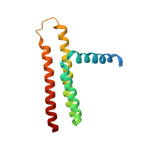
    Membrane proteins play vital roles in the life of the cell and are important therapeutic targets. Producing them in large quantities, pure and fully functional is a major challenge. Many promising projects end when intractable aggregates or precipitates form. Here we show how such unfolded aggregates can be solubilized and the solution mixed with lipid to spontaneously self-assemble a bicontinuous cubic mesophase into the bilayer of which the protein, in a confined, chaperonin-like environment, reconstitutes with 100% efficiency. The test protein, diacylglycerol kinase, reconstituted in the bilayer of the mesophase, was then crystallized in situ by the in meso or lipid cubic phase method providing an X-ray structure to a resolution of 2.55 Å. This highly efficient, inexpensive, simple and rapid approach should find application wherever properly folded, membrane reconstituted and functional proteins are required where the starting material is a denatured aggregate.


  • Organizational Affiliation

    School of Biochemistry and Immunology & School of Medicine, Trinity College Dublin, Dublin, Ireland.


Macromolecules
Find similar proteins by:  (by identity cutoff)  |  3D Structure
Entity ID: 1
MoleculeChains Sequence LengthOrganismDetailsImage
DIACYLGLYCEROL KINASE
A, B, C
130Escherichia coli K-12Mutation(s): 0 
EC: 2.7.1.107
Membrane Entity: Yes 
UniProt
Find proteins for P0ABN1 (Escherichia coli (strain K12))
Explore P0ABN1 
Go to UniProtKB:  P0ABN1
Entity Groups  
Sequence Clusters30% Identity50% Identity70% Identity90% Identity95% Identity100% Identity
UniProt GroupP0ABN1
Sequence Annotations
Expand
  • Reference Sequence
Experimental Data & Validation

Experimental Data

  • Method: X-RAY DIFFRACTION
  • Resolution: 3.80 Å
  • R-Value Free: 0.371 
  • R-Value Work: 0.325 
  • R-Value Observed: 0.326 
  • Space Group: P 31 2 1
Unit Cell:
Length ( Å )Angle ( ˚ )
a = 75.39α = 90
b = 75.39β = 90
c = 197.2γ = 120
Software Package:
Software NamePurpose
PHENIXrefinement
XDSdata reduction
XSCALEdata scaling
PHASERphasing

Structure Validation

View Full Validation Report


Entry History 

Deposition Data

Revision History  (Full details and data files)

  • Version 1.0: 2014-10-22
    Type: Initial release
  • Version 1.1: 2019-03-06
    Changes: Data collection, Experimental preparation, Other
  • Version 1.2: 2019-04-03
    Changes: Data collection, Experimental preparation
  • Version 1.3: 2024-01-10
    Changes: Data collection, Database references, Other, Refinement description