4V40

BETA-GALACTOSIDASE


Experimental Data Snapshot

  • Method: X-RAY DIFFRACTION
  • Resolution: 2.50 Å
  • R-Value Observed: 0.174 

wwPDB Validation   3D Report Full Report


This is version 1.2 of the entry. See complete history


Literature

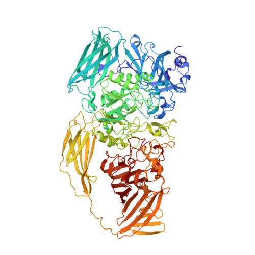
Three-dimensional structure of beta-galactosidase from E. coli.

Jacobson, R.H.Zhang, X.J.DuBose, R.F.Matthews, B.W.

(1994) Nature 369: 761-766

  • DOI: https://doi.org/10.1038/369761a0
  • Primary Citation of Related Structures:  
    4V40

  • PubMed Abstract: 

    The beta-galactosidase from Escherichia coli was instrumental in the development of the operon model, and today is one of the most commonly used enzymes in molecular biology. Here we report the structure of this protein and show that it is a tetramer with 222-point symmetry. The 1,023-amino-acid polypeptide chain folds into five sequential domains, with an extended segment at the amino terminus. The participation of this amino-terminal segment in a subunit interface, coupled with the observation that each active site is made up of elements from two different subunits, provides a structural rationale for the phenomenon of alpha-complementation. The structure represents the longest polypeptide chain for which an atomic structure has been determined. Our results show that it is possible successfully to study non-viral protein crystals with unit cell dimensions in excess of 500 A and with relative molecular masses in the region of 2,000K per asymmetric unit. Non-crystallographic symmetry averaging proved to be a very powerful tool in the structure determination, as has been shown in other contexts.


  • Organizational Affiliation

    Institute of Molecular Biology, Howard Hughes Medical Institute, University of Oregon, Eugene 97403.


Macromolecules
Find similar proteins by:  (by identity cutoff)  |  3D Structure
Entity ID: 1
MoleculeChains Sequence LengthOrganismDetailsImage
BETA-GALACTOSIDASE
A, B, C, D, E
A, B, C, D, E, F, G, H, I, J, K, L, M, N, O, P
1,023Escherichia coliMutation(s): 0 
EC: 3.2.1.23
UniProt
Find proteins for P00722 (Escherichia coli (strain K12))
Explore P00722 
Go to UniProtKB:  P00722
Entity Groups  
Sequence Clusters30% Identity50% Identity70% Identity90% Identity95% Identity100% Identity
UniProt GroupP00722
Sequence Annotations
Expand
  • Reference Sequence
Small Molecules
Ligands 1 Unique
IDChains Name / Formula / InChI Key2D Diagram3D Interactions
MG
Query on MG

Download Ideal Coordinates CCD File 
AA [auth F]
BA [auth F]
CA [auth G]
DA [auth G]
EA [auth H]
AA [auth F],
BA [auth F],
CA [auth G],
DA [auth G],
EA [auth H],
FA [auth H],
GA [auth I],
HA [auth I],
IA [auth J],
JA [auth J],
KA [auth K],
LA [auth K],
MA [auth L],
NA [auth L],
OA [auth M],
PA [auth N],
Q [auth A],
QA [auth N],
R [auth A],
RA [auth O],
S [auth B],
SA [auth O],
T [auth B],
TA [auth P],
U [auth C],
UA [auth P],
V [auth C],
W [auth D],
X [auth D],
Y [auth E],
Z [auth E]
MAGNESIUM ION
Mg
JLVVSXFLKOJNIY-UHFFFAOYSA-N
Experimental Data & Validation

Experimental Data

  • Method: X-RAY DIFFRACTION
  • Resolution: 2.50 Å
  • R-Value Observed: 0.174 
  • Space Group: P 1 21 1
Unit Cell:
Length ( Å )Angle ( ˚ )
a = 107.9α = 90
b = 207.5β = 94.7
c = 509.9γ = 90
Software Package:
Software NamePurpose
TNTrefinement
TNTrefinement
WEISdata reduction

Structure Validation

View Full Validation Report


Entry History 

Deposition Data

Revision History  (Full details and data files)

  • Version 1.0: 2014-07-09
    Type: Initial release
  • Version 1.1: 2014-12-10
    Changes: Other
  • Version 1.2: 2024-02-28
    Changes: Data collection, Database references, Derived calculations, Other