4YCN

Crystal structure of the calcium pump with bound marine macrolide BLLB


Experimental Data Snapshot

  • Method: X-RAY DIFFRACTION
  • Resolution: 3.50 Å
  • R-Value Free: 0.247 
  • R-Value Work: 0.210 
  • R-Value Observed: 0.211 

wwPDB Validation   3D Report Full Report


Ligand Structure Quality Assessment 


This is version 1.1 of the entry. See complete history


Literature

Biselyngbyasides, cytotoxic marine macrolides, are novel and potent inhibitors of the Ca(2+) pumps with a unique mode of binding

Morita, M.Ogawa, H.Ohno, O.Yamori, T.Suenaga, K.Toyoshima, C.

(2015) FEBS Lett 589: 1406-1411

  • DOI: https://doi.org/10.1016/j.febslet.2015.04.056
  • Primary Citation of Related Structures:  
    4YCM, 4YCN

  • PubMed Abstract: 

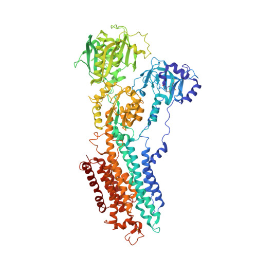
    Biselyngbyasides (BLSs), macrolides from a marine cyanobacterium, are cytotoxic natural products whose target molecule is unknown. Here we report that BLSs are high affinity (Ki∼10 nM) inhibitors of Ca(2+)-pumps with a unique binding mode. The crystal structures of the Ca(2+)-pump in complex with BLSs at 3.2-3.5 Å-resolution show that BLSs bind to the pump near the cytoplasmic surface of the transmembrane region. The crystal structures and activity measurement of BLS analogs allow us to identify the structural features that confer high potency to BLSs as inhibitors of the pump.


  • Organizational Affiliation

    Department of Chemistry, Keio University, Kohoku-ku, Yokohama, Kanagawa 223-8522, Japan; Institute of Molecular and Cellular Biosciences, The University of Tokyo, Bunkyo-ku, Tokyo 113-0032, Japan.


Macromolecules
Find similar proteins by:  (by identity cutoff)  |  3D Structure
Entity ID: 1
MoleculeChains Sequence LengthOrganismDetailsImage
Sarcoplasmic/endoplasmic reticulum calcium ATPase 1994Oryctolagus cuniculusMutation(s): 0 
EC: 3.6.3.8
Membrane Entity: Yes 
UniProt
Find proteins for P04191 (Oryctolagus cuniculus)
Explore P04191 
Go to UniProtKB:  P04191
Entity Groups  
Sequence Clusters30% Identity50% Identity70% Identity90% Identity95% Identity100% Identity
UniProt GroupP04191
Sequence Annotations
Expand
  • Reference Sequence
Small Molecules
Ligands 4 Unique
IDChains Name / Formula / InChI Key2D Diagram3D Interactions
PTY
Query on PTY

Download Ideal Coordinates CCD File 
F [auth A],
G [auth A]
PHOSPHATIDYLETHANOLAMINE
C40 H80 N O8 P
NJGIRBISCGPRPF-KXQOOQHDSA-N
7BL
Query on 7BL

Download Ideal Coordinates CCD File 
E [auth A](4S,5E,8S,9E,11S,13E,15E,18R)-4-hydroxy-8-methoxy-9,11-dimethyl-18-[(1Z,4E)-2-methylhexa-1,4-dien-1-yl]oxacyclooctadeca-5,9,13,15-tetraen-2-one
C27 H40 O4
DVYYXLNVAXSASA-BEVCVMOBSA-N
SO4
Query on SO4

Download Ideal Coordinates CCD File 
C [auth A],
D [auth A]
SULFATE ION
O4 S
QAOWNCQODCNURD-UHFFFAOYSA-L
NA
Query on NA

Download Ideal Coordinates CCD File 
B [auth A]SODIUM ION
Na
FKNQFGJONOIPTF-UHFFFAOYSA-N
Binding Affinity Annotations 
IDSourceBinding Affinity
7BL BindingDB:  4YCN Ki: min: 17, max: 17 (nM) from 2 assay(s)
Experimental Data & Validation

Experimental Data

  • Method: X-RAY DIFFRACTION
  • Resolution: 3.50 Å
  • R-Value Free: 0.247 
  • R-Value Work: 0.210 
  • R-Value Observed: 0.211 
  • Space Group: P 41 21 2
Unit Cell:
Length ( Å )Angle ( ˚ )
a = 71.54α = 90
b = 71.54β = 90
c = 587.669γ = 90
Software Package:
Software NamePurpose
PHENIXrefinement
DENZOdata scaling
SCALEPACKdata scaling

Structure Validation

View Full Validation Report


Ligand Structure Quality Assessment 


Entry History 

Deposition Data

Revision History  (Full details and data files)

  • Version 1.0: 2016-01-13
    Type: Initial release
  • Version 1.1: 2020-02-05
    Changes: Data collection, Database references, Derived calculations