5CGB

Crystal structure of FimH in complex with heptyl alpha-D-septanoside


Experimental Data Snapshot

  • Method: X-RAY DIFFRACTION
  • Resolution: 1.60 Å
  • R-Value Free: 0.179 
  • R-Value Work: 0.160 
  • R-Value Observed: 0.161 

wwPDB Validation   3D Report Full Report


Ligand Structure Quality Assessment 


This is version 1.2 of the entry. See complete history


Literature

The price of flexibility - a case study on septanoses as pyranose mimetics.

Sager, C.P.Fiege, B.Zihlmann, P.Vannam, R.Rabbani, S.Jakob, R.P.Preston, R.C.Zalewski, A.Maier, T.Peczuh, M.W.Ernst, B.

(2018) Chem Sci 9: 646-654

  • DOI: https://doi.org/10.1039/c7sc04289b
  • Primary Citation of Related Structures:  
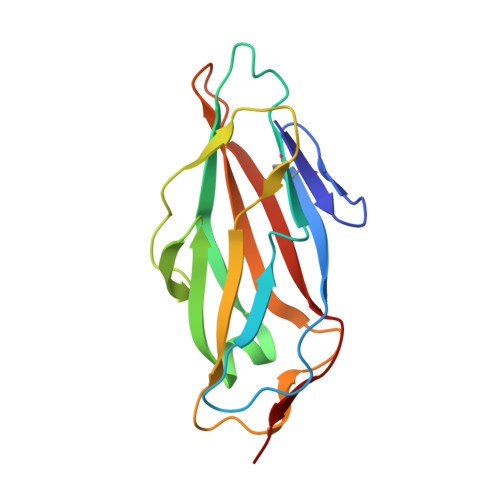
    5CGB

  • PubMed Abstract: 

    Seven-membered ring mimetics of mannose were studied as ligands for the mannose-specific bacterial lectin FimH, which plays an essential role in the first step of urinary tract infections (UTI). A competitive binding assay and isothermal titration calorimetry (ITC) experiments indicated an approximately ten-fold lower affinity for the seven-membered ring mannose mimetic 2- O-n -heptyl-1,6-anhydro-d- glycero -d-galactitol ( 7 ) compared to n -heptyl α-d-mannopyranoside ( 2 ), resulting exclusively from a loss of conformational entropy. Investigations by solution NMR, X-ray crystallography, and molecular modeling revealed that 7 establishes a superimposable H-bond network compared to mannoside 2 , but at the price of a high entropic penalty due to the loss of its pronounced conformational flexibility. These results underscore the importance of having access to the complete thermodynamic profile of a molecular interaction to "rescue" ligands from entropic penalties with an otherwise perfect fit to the protein binding site.


  • Organizational Affiliation

    University of Basel , Institute of Molecular Pharmacy , Pharmacenter of the University of Basel , Klingelbergstrasse 50 , 4056 , Basel , Switzerland . Email: beat.ernst@unibas.ch.


Macromolecules
Find similar proteins by:  (by identity cutoff)  |  3D Structure
Entity ID: 1
MoleculeChains Sequence LengthOrganismDetailsImage
Protein FimH
A, B
158Escherichia coli K-12Mutation(s): 0 
Gene Names: fimHb4320JW4283
UniProt
Find proteins for P08191 (Escherichia coli (strain K12))
Explore P08191 
Go to UniProtKB:  P08191
Entity Groups  
Sequence Clusters30% Identity50% Identity70% Identity90% Identity95% Identity100% Identity
UniProt GroupP08191
Sequence Annotations
Expand
  • Reference Sequence
Experimental Data & Validation

Experimental Data

  • Method: X-RAY DIFFRACTION
  • Resolution: 1.60 Å
  • R-Value Free: 0.179 
  • R-Value Work: 0.160 
  • R-Value Observed: 0.161 
  • Space Group: P 21 21 21
Unit Cell:
Length ( Å )Angle ( ˚ )
a = 63.58α = 90
b = 68.55β = 90
c = 95.78γ = 90
Software Package:
Software NamePurpose
PHENIXrefinement
XDSdata reduction
XDSdata scaling
PHASERphasing

Structure Validation

View Full Validation Report


Ligand Structure Quality Assessment 


Entry History 

Deposition Data

Revision History  (Full details and data files)

  • Version 1.0: 2016-07-20
    Type: Initial release
  • Version 1.1: 2021-04-07
    Changes: Database references
  • Version 1.2: 2024-01-10
    Changes: Data collection, Database references, Refinement description