5DSV

Crystal structure of human proteasome alpha7 tetradecamer


Experimental Data Snapshot

  • Method: X-RAY DIFFRACTION
  • Resolution: 3.75 Å
  • R-Value Free: 0.291 
  • R-Value Work: 0.209 
  • R-Value Observed: 0.213 

wwPDB Validation   3D Report Full Report


This is version 1.2 of the entry. See complete history


Literature

Disassembly of the self-assembled, double-ring structure of proteasome alpha 7 homo-tetradecamer by alpha 6

Ishii, K.Noda, M.Yagi, H.Thammaporn, R.Seetaha, S.Satoh, T.Kato, K.Uchiyama, S.

(2015) Sci Rep 5: 18167-18167

  • DOI: https://doi.org/10.1038/srep18167
  • Primary Citation of Related Structures:  
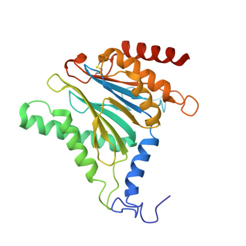
    5DSV

  • PubMed Abstract: 

    The 20S core particle of the eukaryotic proteasome is composed of two α- and two β-rings, each of which is a hetero-heptamer composed of seven homologous but distinct subunits. Although formation of the eukaryotic proteasome is a highly ordered process assisted by assembly chaperones, α7, an α-ring component, has the unique property of self-assembling into a homo-tetradecamer. We used biophysical methods to characterize the oligomeric states of this proteasome subunit and its interaction with α6, which makes direct contacts with α7 in the proteasome α-ring. We determined a crystal structure of the α7 tetradecamer, which has a double-ring structure. Sedimentation velocity analytical ultracentrifugation and mass spectrometric analysis under non-denaturing conditions revealed that α7 exclusively exists as homo-tetradecamer in solution and that its double-ring structure is disassembled upon the addition of α6, resulting in a 1:7 hetero-octameric α6-α7 complex. Our findings suggest that proteasome formation involves the disassembly of non-native oligomers, which are assembly intermediates.


  • Organizational Affiliation

    Okazaki Institute for Integrative Bioscience, National Institutes of Natural Sciences, 5-1 Higashiyama, Myodaiji, Okazaki, Aichi 444-8787, Japan.


Macromolecules
Find similar proteins by:  (by identity cutoff)  |  3D Structure
Entity ID: 1
MoleculeChains Sequence LengthOrganismDetailsImage
Proteasome subunit alpha type-3
A, B, C, D, E
A, B, C, D, E, F, G, H, I, J, K, L, M, N
255Homo sapiensMutation(s): 0 
Gene Names: PSMA3HC8PSC8
EC: 3.4.25.1
UniProt & NIH Common Fund Data Resources
Find proteins for P25788 (Homo sapiens)
Explore P25788 
Go to UniProtKB:  P25788
PHAROS:  P25788
GTEx:  ENSG00000100567 
Entity Groups  
Sequence Clusters30% Identity50% Identity70% Identity90% Identity95% Identity100% Identity
UniProt GroupP25788
Sequence Annotations
Expand
  • Reference Sequence
Experimental Data & Validation

Experimental Data

  • Method: X-RAY DIFFRACTION
  • Resolution: 3.75 Å
  • R-Value Free: 0.291 
  • R-Value Work: 0.209 
  • R-Value Observed: 0.213 
  • Space Group: P 43 21 2
Unit Cell:
Length ( Å )Angle ( ˚ )
a = 132.481α = 90
b = 132.481β = 90
c = 444.563γ = 90
Software Package:
Software NamePurpose
REFMACrefinement
SCALEPACKdata reduction
DENZOdata scaling
MOLREPphasing

Structure Validation

View Full Validation Report


Entry History & Funding Information

Deposition Data


Funding OrganizationLocationGrant Number
JSPS, MEXT, JapanJapan25102001, 25102008, and 15H02491 to K.K. and 25121722 and 26102530 to S.U.

Revision History  (Full details and data files)

  • Version 1.0: 2015-12-02
    Type: Initial release
  • Version 1.1: 2015-12-30
    Changes: Database references
  • Version 1.2: 2023-11-08
    Changes: Data collection, Database references, Derived calculations, Refinement description