5EE4

The crystal structure of HpuA from Kingella denitrificans in complex with human haemoglobin


Experimental Data Snapshot

  • Method: X-RAY DIFFRACTION
  • Resolution: 2.30 Å
  • R-Value Free: 0.244 
  • R-Value Work: 0.202 
  • R-Value Observed: 0.204 

wwPDB Validation   3D Report Full Report


Ligand Structure Quality Assessment 


This is version 1.3 of the entry. See complete history


Literature

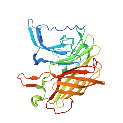
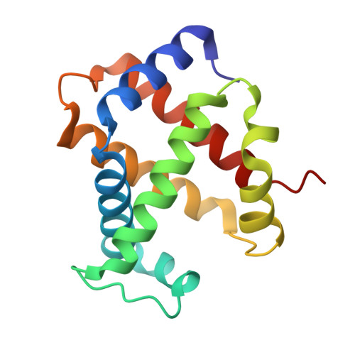
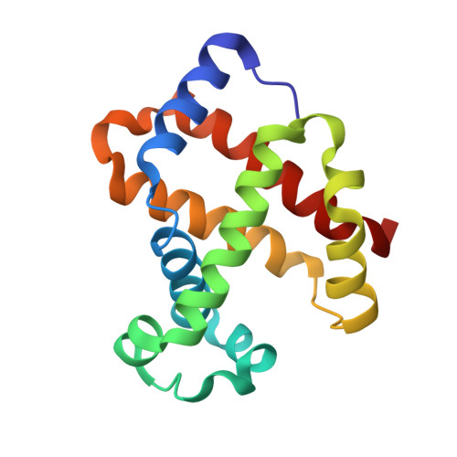
Structural analysis of haemoglobin binding by HpuA from the Neisseriaceae family.

Wong, C.T.Xu, Y.Gupta, A.Garnett, J.A.Matthews, S.J.Hare, S.A.

(2015) Nat Commun 6: 10172-10172

  • DOI: https://doi.org/10.1038/ncomms10172
  • Primary Citation of Related Structures:  
    5EC6, 5EE2, 5EE4

  • PubMed Abstract: 

    The Neisseriaceae family of bacteria causes a range of diseases including meningitis, septicaemia, gonorrhoea and endocarditis, and extracts haem from haemoglobin as an important iron source within the iron-limited environment of its human host. Herein we report crystal structures of apo- and haemoglobin-bound HpuA, an essential component of this haem import system. The interface involves long loops on the bacterial receptor that present hydrophobic side chains for packing against the surface of haemoglobin. Interestingly, our structural and biochemical analyses of Kingella denitrificans and Neisseria gonorrhoeae HpuA mutants, although validating the interactions observed in the crystal structure, show how Neisseriaceae have the fascinating ability to diversify functional sequences and yet retain the haemoglobin binding function. Our results present the first description of HpuA's role in direct binding of haemoglobin.


  • Organizational Affiliation

    Department of Life Sciences, Imperial College London, South Kensington Campus, Exhibition Road, London SW7 2AZ, UK.


Macromolecules
Find similar proteins by:  (by identity cutoff)  |  3D Structure
Entity ID: 1
MoleculeChains Sequence LengthOrganismDetailsImage
HpuA
A, B
322Kingella denitrificans ATCC 33394Mutation(s): 0 
Gene Names: HMPREF9098_0447
UniProt
Find proteins for F0EX68 (Kingella denitrificans ATCC 33394)
Explore F0EX68 
Go to UniProtKB:  F0EX68
Entity Groups  
Sequence Clusters30% Identity50% Identity70% Identity90% Identity95% Identity100% Identity
UniProt GroupF0EX68
Sequence Annotations
Expand
  • Reference Sequence
Find similar proteins by:  (by identity cutoff)  |  3D Structure
Entity ID: 2
MoleculeChains Sequence LengthOrganismDetailsImage
Hemoglobin subunit alpha
C, E
141Homo sapiensMutation(s): 0 
UniProt & NIH Common Fund Data Resources
Find proteins for P69905 (Homo sapiens)
Explore P69905 
Go to UniProtKB:  P69905
PHAROS:  P69905
Entity Groups  
Sequence Clusters30% Identity50% Identity70% Identity90% Identity95% Identity100% Identity
UniProt GroupP69905
Sequence Annotations
Expand
  • Reference Sequence
Find similar proteins by:  (by identity cutoff)  |  3D Structure
Entity ID: 3
MoleculeChains Sequence LengthOrganismDetailsImage
Hemoglobin subunit beta
D, F
146Homo sapiensMutation(s): 0 
UniProt & NIH Common Fund Data Resources
Find proteins for P68871 (Homo sapiens)
Explore P68871 
Go to UniProtKB:  P68871
PHAROS:  P68871
GTEx:  ENSG00000244734 
Entity Groups  
Sequence Clusters30% Identity50% Identity70% Identity90% Identity95% Identity100% Identity
UniProt GroupP68871
Sequence Annotations
Expand
  • Reference Sequence
Small Molecules
Ligands 3 Unique
IDChains Name / Formula / InChI Key2D Diagram3D Interactions
HEM
Query on HEM

Download Ideal Coordinates CCD File 
H [auth C],
J [auth D],
M [auth E],
O [auth F]
PROTOPORPHYRIN IX CONTAINING FE
C34 H32 Fe N4 O4
KABFMIBPWCXCRK-RGGAHWMASA-L
GOL
Query on GOL

Download Ideal Coordinates CCD File 
G [auth A],
L [auth D]
GLYCEROL
C3 H8 O3
PEDCQBHIVMGVHV-UHFFFAOYSA-N
OXY
Query on OXY

Download Ideal Coordinates CCD File 
I [auth C],
K [auth D],
N [auth E],
P [auth F]
OXYGEN MOLECULE
O2
MYMOFIZGZYHOMD-UHFFFAOYSA-N
Experimental Data & Validation

Experimental Data

  • Method: X-RAY DIFFRACTION
  • Resolution: 2.30 Å
  • R-Value Free: 0.244 
  • R-Value Work: 0.202 
  • R-Value Observed: 0.204 
  • Space Group: P 1 21 1
Unit Cell:
Length ( Å )Angle ( ˚ )
a = 54.79α = 90
b = 87.21β = 98.13
c = 124.32γ = 90
Software Package:
Software NamePurpose
MOSFLMdata reduction
SCALAdata scaling
PHASERphasing
REFMACrefinement
PDB_EXTRACTdata extraction

Structure Validation

View Full Validation Report


Ligand Structure Quality Assessment 


Entry History & Funding Information

Deposition Data


Funding OrganizationLocationGrant Number
Medical Research Council (United Kingdom)United KingdomG1100332

Revision History  (Full details and data files)

  • Version 1.0: 2015-11-04
    Type: Initial release
  • Version 1.1: 2015-12-30
    Changes: Database references
  • Version 1.2: 2017-09-13
    Changes: Author supporting evidence, Data collection
  • Version 1.3: 2024-01-10
    Changes: Data collection, Database references, Derived calculations, Refinement description