5GSE

Crystal structure of unusual nucleosome


Experimental Data Snapshot

  • Method: X-RAY DIFFRACTION
  • Resolution: 3.14 Å
  • R-Value Free: 0.255 
  • R-Value Work: 0.197 
  • R-Value Observed: 0.200 

wwPDB Validation   3D Report Full Report


This is version 1.1 of the entry. See complete history


Literature

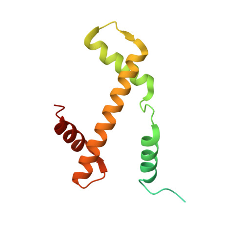
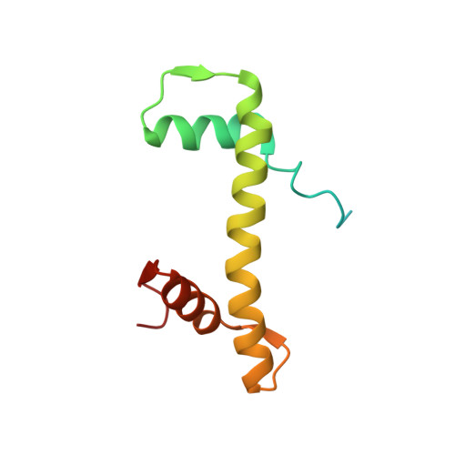
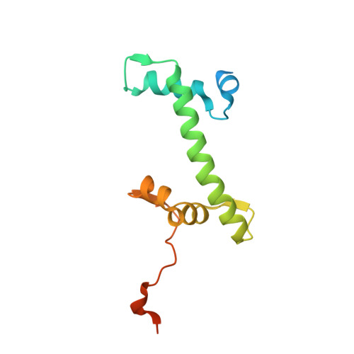
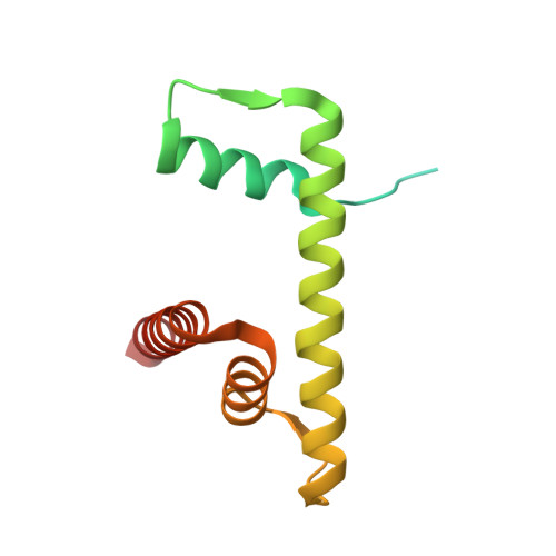
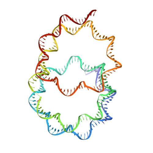
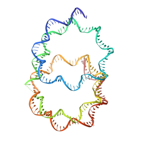
Crystal structure of the overlapping dinucleosome composed of hexasome and octasome

Kato, D.Osakabe, A.Arimura, Y.Mizukami, Y.Horikoshi, N.Saikusa, K.Akashi, S.Nishimura, Y.Park, S.Y.Nogami, J.Maehara, K.Ohkawa, Y.Matsumoto, A.Kono, H.Inoue, R.Sugiyama, M.Kurumizaka, H.

(2017) Science 356: 205-208

  • DOI: https://doi.org/10.1126/science.aak9867
  • Primary Citation of Related Structures:  
    5GSE

  • PubMed Abstract: 

    Nucleosomes are dynamic entities that are repositioned along DNA by chromatin remodeling processes. A nucleosome repositioned by the switch-sucrose nonfermentable (SWI/SNF) remodeler collides with a neighbor and forms the intermediate "overlapping dinucleosome." Here, we report the crystal structure of the overlapping dinucleosome, in which two nucleosomes are associated, at 3.14-angstrom resolution. In the overlapping dinucleosome structure, the unusual "hexasome" nucleosome, composed of the histone hexamer lacking one H2A-H2B dimer from the conventional histone octamer, contacts the canonical "octasome" nucleosome, and they intimately associate. Consequently, about 250 base pairs of DNA are left-handedly wrapped in three turns, without a linker DNA segment between the hexasome and octasome moieties. The overlapping dinucleosome structure may provide important information to understand how nucleosome repositioning occurs during the chromatin remodeling process.


  • Organizational Affiliation

    Laboratory of Structural Biology, Graduate School of Advanced Science and Engineering, Waseda University, 2-2 Wakamatsu-cho, Shinjuku-ku, Tokyo 162-8480, Japan.


Macromolecules

Find similar proteins by:  (by identity cutoff)  |  3D Structure
Entity ID: 1
MoleculeChains Sequence LengthOrganismDetailsImage
Histone H3.1
A, E, K, O
139Homo sapiensMutation(s): 0 
Gene Names: HIST1H3A
UniProt & NIH Common Fund Data Resources
Find proteins for P68431 (Homo sapiens)
Explore P68431 
Go to UniProtKB:  P68431
PHAROS:  P68431
Entity Groups  
Sequence Clusters30% Identity50% Identity70% Identity90% Identity95% Identity100% Identity
UniProt GroupP68431
Sequence Annotations
Expand
  • Reference Sequence
Find similar proteins by:  (by identity cutoff)  |  3D Structure
Entity ID: 2
MoleculeChains Sequence LengthOrganismDetailsImage
Histone H4
B, F, L, P
106Homo sapiensMutation(s): 0 
Gene Names: HIST1H4A
UniProt & NIH Common Fund Data Resources
Find proteins for P62805 (Homo sapiens)
Explore P62805 
Go to UniProtKB:  P62805
PHAROS:  P62805
Entity Groups  
Sequence Clusters30% Identity50% Identity70% Identity90% Identity95% Identity100% Identity
UniProt GroupP62805
Sequence Annotations
Expand
  • Reference Sequence
Find similar proteins by:  (by identity cutoff)  |  3D Structure
Entity ID: 3
MoleculeChains Sequence LengthOrganismDetailsImage
Histone H2A type 1-B/E
C, G, M
133Homo sapiensMutation(s): 0 
Gene Names: HIST1H2AB
UniProt & NIH Common Fund Data Resources
Find proteins for P04908 (Homo sapiens)
Explore P04908 
Go to UniProtKB:  P04908
PHAROS:  P04908
Entity Groups  
Sequence Clusters30% Identity50% Identity70% Identity90% Identity95% Identity100% Identity
UniProt GroupP04908
Sequence Annotations
Expand
  • Reference Sequence
Find similar proteins by:  (by identity cutoff)  |  3D Structure
Entity ID: 4
MoleculeChains Sequence LengthOrganismDetailsImage
Histone H2B type 1-J
D, H, N
129Homo sapiensMutation(s): 0 
Gene Names: HIST1H2BJ
UniProt & NIH Common Fund Data Resources
Find proteins for P06899 (Homo sapiens)
Explore P06899 
Go to UniProtKB:  P06899
PHAROS:  P06899
GTEx:  ENSG00000124635 
Entity Groups  
Sequence Clusters30% Identity50% Identity70% Identity90% Identity95% Identity100% Identity
UniProt GroupP06899
Sequence Annotations
Expand
  • Reference Sequence
Find similar nucleic acids by:  (by identity cutoff)  |  3D Structure
Entity ID: 5
MoleculeChains LengthOrganismImage
DNA (250-MER)250synthetic construct
Sequence Annotations
Expand
  • Reference Sequence
Find similar nucleic acids by:  (by identity cutoff)  |  3D Structure
Entity ID: 6
MoleculeChains LengthOrganismImage
DNA (250-MER)250synthetic construct
Sequence Annotations
Expand
  • Reference Sequence
Experimental Data & Validation

Experimental Data

  • Method: X-RAY DIFFRACTION
  • Resolution: 3.14 Å
  • R-Value Free: 0.255 
  • R-Value Work: 0.197 
  • R-Value Observed: 0.200 
  • Space Group: P 1
Unit Cell:
Length ( Å )Angle ( ˚ )
a = 90.582α = 119.3
b = 101.804β = 106.51
c = 102.427γ = 91.36
Software Package:
Software NamePurpose
PHENIXrefinement
HKL-2000data collection
HKL-2000data scaling
MOLREPphasing
Cootmodel building
PDB_EXTRACTdata extraction

Structure Validation

View Full Validation Report


Entry History & Funding Information

Deposition Data


Funding OrganizationLocationGrant Number
MEXT KAKENHI GrantJapan25116002
Agency for Medical Research and Development, the Platform Project for Supporting Drug Discovery and Life Science ResearchJapan--
the Waseda Research Institute for Science and EngineeringJapan--
Japan Society for the Promotion of ScienceJapan15H06691
Japan Society for the Promotion of ScienceJapan16J06850

Revision History  (Full details and data files)

  • Version 1.0: 2017-05-03
    Type: Initial release
  • Version 1.1: 2023-11-08
    Changes: Data collection, Database references, Refinement description