5KKE

Crystal Structure of a Domain-swapped Dimer of Yeast Iso-1-cytochrome c with CYMAL5


Experimental Data Snapshot

  • Method: X-RAY DIFFRACTION
  • Resolution: 1.70 Å
  • R-Value Free: 0.204 
  • R-Value Work: 0.171 
  • R-Value Observed: 0.174 

wwPDB Validation   3D Report Full Report


Ligand Structure Quality Assessment 


This is version 2.0 of the entry. See complete history


Literature

Cytochrome c Can Form a Well-Defined Binding Pocket for Hydrocarbons.

McClelland, L.J.Steele, H.B.Whitby, F.G.Mou, T.C.Holley, D.Ross, J.B.Sprang, S.R.Bowler, B.E.

(2016) J Am Chem Soc 138: 16770-16778

  • DOI: https://doi.org/10.1021/jacs.6b10745
  • Primary Citation of Related Structures:  
    5KKE, 5KLU, 5T7H

  • PubMed Abstract: 

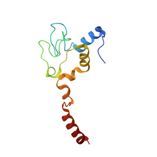
    Cytochrome c can acquire peroxidase activity when it binds to cardiolipin in mitochondrial membranes. The resulting oxygenation of cardiolipin by cytochrome c provides an early signal for the onset of apoptosis. The structure of this enzyme-substrate complex is a matter of considerable debate. We present three structures at 1.7-2.0 Å resolution of a domain-swapped dimer of yeast iso-1-cytochrome c with the detergents, CYMAL-5, CYMAL-6, and ω-undecylenyl-β-d-maltopyranoside, bound in a channel that places the hydrocarbon moieties of these detergents next to the heme. The heme is poised for peroxidase activity with water bound in place of Met80, which serves as the axial heme ligand when cytochrome c functions as an electron carrier. The hydroxyl group of Tyr67 sits 3.6-4.0 Å from the nearest carbon of the detergents, positioned to act as a relay in radical abstraction during peroxidase activity. Docking studies with linoleic acid, the most common fatty acid component of cardiolipin, show that C11 of linoleic acid can sit adjacent to Tyr67 and the heme, consistent with the oxygenation pattern observed in lipidomics studies. The well-defined hydrocarbon binding pocket provides atomic resolution evidence for the extended lipid anchorage model for cytochrome c/cardiolipin binding. Dimer dissociation/association kinetics for yeast versus equine cytochrome c indicate that formation of mammalian cytochrome c dimers in vivo would require catalysis. However, the dimer structure shows that only a modest deformation of monomeric cytochrome c would suffice to form the hydrocarbon binding site occupied by these detergents.


  • Organizational Affiliation

    Department of Chemistry & Biochemistry, University of Montana , Missoula, Montana 59812, United States.


Macromolecules
Find similar proteins by:  (by identity cutoff)  |  3D Structure
Entity ID: 1
MoleculeChains Sequence LengthOrganismDetailsImage
Cytochrome c iso-1108Saccharomyces cerevisiae S288CMutation(s): 2 
Gene Names: CYC1YJR048WJ1653
UniProt
Find proteins for P00044 (Saccharomyces cerevisiae (strain ATCC 204508 / S288c))
Explore P00044 
Go to UniProtKB:  P00044
Entity Groups  
Sequence Clusters30% Identity50% Identity70% Identity90% Identity95% Identity100% Identity
UniProt GroupP00044
Sequence Annotations
Expand
  • Reference Sequence
Small Molecules
Ligands 5 Unique
IDChains Name / Formula / InChI Key2D Diagram3D Interactions
HEC
Query on HEC

Download Ideal Coordinates CCD File 
B [auth A]HEME C
C34 H34 Fe N4 O4
HXQIYSLZKNYNMH-LJNAALQVSA-N
CM5
Query on CM5

Download Ideal Coordinates CCD File 
E [auth A]5-CYCLOHEXYL-1-PENTYL-BETA-D-MALTOSIDE
C23 H42 O11
RVTGFZGNOSKUDA-ZNGNCRBCSA-N
SO4
Query on SO4

Download Ideal Coordinates CCD File 
C [auth A],
D [auth A]
SULFATE ION
O4 S
QAOWNCQODCNURD-UHFFFAOYSA-L
GOL
Query on GOL

Download Ideal Coordinates CCD File 
G [auth A],
H [auth A]
GLYCEROL
C3 H8 O3
PEDCQBHIVMGVHV-UHFFFAOYSA-N
CL
Query on CL

Download Ideal Coordinates CCD File 
F [auth A]CHLORIDE ION
Cl
VEXZGXHMUGYJMC-UHFFFAOYSA-M
Experimental Data & Validation

Experimental Data

  • Method: X-RAY DIFFRACTION
  • Resolution: 1.70 Å
  • R-Value Free: 0.204 
  • R-Value Work: 0.171 
  • R-Value Observed: 0.174 
  • Space Group: I 2 2 2
Unit Cell:
Length ( Å )Angle ( ˚ )
a = 49.74α = 90
b = 69.326β = 90
c = 72.482γ = 90
Software Package:
Software NamePurpose
PHENIXrefinement
CrystalCleardata collection
SCALEPACKdata scaling
PHASERphasing
SOLVEphasing
PDB_EXTRACTdata extraction
HKL-2000data reduction

Structure Validation

View Full Validation Report


Ligand Structure Quality Assessment 


Entry History 

Deposition Data

Revision History  (Full details and data files)

  • Version 1.0: 2017-03-22
    Type: Initial release
  • Version 2.0: 2021-03-10
    Changes: Advisory, Atomic model, Data collection, Derived calculations, Non-polymer description, Structure summary