5LZP

Binding of the C-terminal GQYL motif of the bacterial proteasome activator Bpa to the 20S proteasome


Experimental Data Snapshot

  • Method: ELECTRON MICROSCOPY
  • Resolution: 3.50 Å
  • Aggregation State: PARTICLE 
  • Reconstruction Method: SINGLE PARTICLE 

wwPDB Validation   3D Report Full Report


This is version 1.5 of the entry. See complete history


Literature

Structural Analysis of the Bacterial Proteasome Activator Bpa in Complex with the 20S Proteasome.

Bolten, M.Delley, C.L.Leibundgut, M.Boehringer, D.Ban, N.Weber-Ban, E.

(2016) Structure 24: 2138-2151

  • DOI: https://doi.org/10.1016/j.str.2016.10.008
  • Primary Citation of Related Structures:  
    5LFJ, 5LFP, 5LFQ, 5LZP

  • PubMed Abstract: 

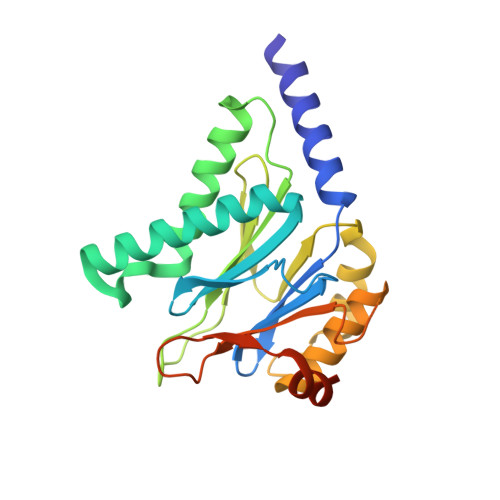
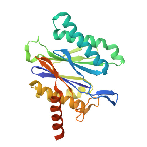
    Mycobacterium tuberculosis harbors proteasomes that recruit substrates for degradation through an ubiquitin-like modification pathway. Recently, a non-ATPase activator termed Bpa (bacterial proteasome activator) was shown to support an alternate proteasomal degradation pathway. Here, we present the cryo-electron microscopy (cryo-EM) structure of Bpa in complex with the 20S core particle (CP). For docking into the cryo-EM density, we solved the X-ray structure of Bpa, showing that it forms tight four-helix bundles arranged into a 12-membered ring with a 40 Å wide central pore and the C-terminal helix of each protomer protruding from the ring. The Bpa model was fitted into the cryo-EM map of the Bpa-CP complex, revealing its architecture and striking symmetry mismatch. The Bpa-CP interface was resolved to 3.5 Å, showing the interactions between the C-terminal GQYL motif of Bpa and the proteasome α-rings. This docking mode is related to the one observed for eukaryotic activators with features specific to the bacterial complex.


  • Organizational Affiliation

    ETH Zurich, Institute of Molecular Biology & Biophysics, 8093 Zurich, Switzerland.


Macromolecules
Find similar proteins by:  (by identity cutoff)  |  3D Structure
Entity ID: 1
MoleculeChains Sequence LengthOrganismDetailsImage
Proteasome subunit alpha241Mycobacterium tuberculosis H37RvMutation(s): 0 
Gene Names: prcARv2109c
EC: 3.4.25.1
UniProt
Find proteins for P9WHU1 (Mycobacterium tuberculosis (strain ATCC 25618 / H37Rv))
Explore P9WHU1 
Go to UniProtKB:  P9WHU1
Entity Groups  
Sequence Clusters30% Identity50% Identity70% Identity90% Identity95% Identity100% Identity
UniProt GroupP9WHU1
Sequence Annotations
Expand
  • Reference Sequence
Find similar proteins by:  (by identity cutoff)  |  3D Structure
Entity ID: 2
MoleculeChains Sequence LengthOrganismDetailsImage
Bacterial proteasome activator180Mycobacterium tuberculosis H37RvMutation(s): 0 
Gene Names: bpaRv3780MTCY13D12.14
UniProt
Find proteins for P9WKX3 (Mycobacterium tuberculosis (strain ATCC 25618 / H37Rv))
Explore P9WKX3 
Go to UniProtKB:  P9WKX3
Entity Groups  
Sequence Clusters30% Identity50% Identity70% Identity90% Identity95% Identity100% Identity
UniProt GroupP9WKX3
Sequence Annotations
Expand
  • Reference Sequence
Find similar proteins by:  (by identity cutoff)  |  3D Structure
Entity ID: 3
MoleculeChains Sequence LengthOrganismDetailsImage
Proteasome subunit beta242Mycobacterium tuberculosis H37RvMutation(s): 0 
Gene Names: prcBRv2110c
EC: 3.4.25.1
UniProt
Find proteins for P9WHT9 (Mycobacterium tuberculosis (strain ATCC 25618 / H37Rv))
Explore P9WHT9 
Go to UniProtKB:  P9WHT9
Entity Groups  
Sequence Clusters30% Identity50% Identity70% Identity90% Identity95% Identity100% Identity
UniProt GroupP9WHT9
Sequence Annotations
Expand
  • Reference Sequence
Experimental Data & Validation

Experimental Data

  • Method: ELECTRON MICROSCOPY
  • Resolution: 3.50 Å
  • Aggregation State: PARTICLE 
  • Reconstruction Method: SINGLE PARTICLE 
EM Software:
TaskSoftware PackageVersion
MODEL REFINEMENTPHENIX1.10-2155
RECONSTRUCTIONRELION1.4

Structure Validation

View Full Validation Report


Entry History 

Deposition Data

Revision History  (Full details and data files)

  • Version 1.0: 2016-11-23
    Type: Initial release
  • Version 1.1: 2016-12-14
    Changes: Database references
  • Version 1.2: 2017-07-26
    Changes: Experimental preparation
  • Version 1.3: 2017-08-02
    Changes: Data collection
  • Version 1.4: 2018-10-17
    Changes: Data collection, Refinement description
  • Version 1.5: 2019-10-23
    Changes: Data collection, Other