5W6G

Human antibody 6649 in complex with influenza hemagglutinin H1 Solomon Islands


Experimental Data Snapshot

  • Method: X-RAY DIFFRACTION
  • Resolution: 2.79 Å
  • R-Value Free: 0.253 
  • R-Value Work: 0.218 
  • R-Value Observed: 0.220 

wwPDB Validation   3D Report Full Report


Ligand Structure Quality Assessment 


This is version 2.1 of the entry. See complete history


Literature

Conserved epitope on influenza-virus hemagglutinin head defined by a vaccine-induced antibody.

Raymond, D.D.Bajic, G.Ferdman, J.Suphaphiphat, P.Settembre, E.C.Moody, M.A.Schmidt, A.G.Harrison, S.C.

(2018) Proc Natl Acad Sci U S A 115: 168-173

  • DOI: https://doi.org/10.1073/pnas.1715471115
  • Primary Citation of Related Structures:  
    5W6C, 5W6G

  • PubMed Abstract: 

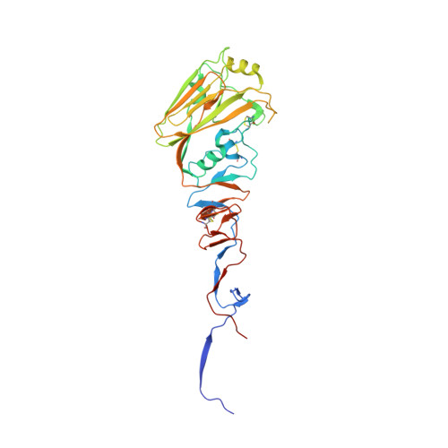
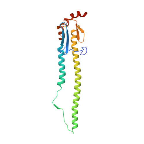
    Circulating influenza viruses evade neutralization in their human hosts by acquiring escape mutations at epitopes of prevalent antibodies. A goal for next-generation influenza vaccines is to reduce escape likelihood by selectively eliciting antibodies recognizing conserved surfaces on the viral hemagglutinin (HA). The receptor-binding site (RBS) on the HA "head" and a region near the fusion peptide on the HA "stem" are two such sites. We describe here a human antibody clonal lineage, designated CL6649, members of which bind a third conserved site ("lateral patch") on the side of the H1-subtype, HA head. A crystal structure of HA with bound Fab6649 shows the conserved antibody footprint. The site was invariant in isolates from 1977 (seasonal) to 2012 (pdm2009); antibodies in CL6649 recognize HAs from the entire period. In 2013, human H1 viruses acquired mutations in this epitope that were retained in subsequent seasons, prompting modification of the H1 vaccine component in 2017. The mutations inhibit Fab6649 binding. We infer from the rapid spread of these mutations in circulating H1 influenza viruses that the previously subdominant, conserved lateral patch had become immunodominant for individuals with B-cell memory imprinted by earlier H1 exposure. We suggest that introduction of the pdm2009 H1 virus, to which most of the broadly prevalent, neutralizing antibodies did not bind, conferred a selective advantage in the immune systems of infected hosts to recall of memory B cells that recognized the lateral patch, the principal exposed epitope that did not change when pdm2009 displaced previous seasonal H1 viruses.


  • Organizational Affiliation

    Laboratory of Molecular Medicine, Boston Children's Hospital, Harvard Medical School, Boston, MA 02115.


Macromolecules
Find similar proteins by:  (by identity cutoff)  |  3D Structure
Entity ID: 1
MoleculeChains Sequence LengthOrganismDetailsImage
Hemagglutinin HA1334Influenza A virus (A/Solomon Islands/3/2006(H1N1))Mutation(s): 0 
Gene Names: HA
UniProt
Find proteins for A7UPX0 (Influenza A virus)
Explore A7UPX0 
Go to UniProtKB:  A7UPX0
Entity Groups  
Sequence Clusters30% Identity50% Identity70% Identity90% Identity95% Identity100% Identity
UniProt GroupA7UPX0
Sequence Annotations
Expand
  • Reference Sequence
Find similar proteins by:  (by identity cutoff)  |  3D Structure
Entity ID: 2
MoleculeChains Sequence LengthOrganismDetailsImage
Hemagglutinin HA2178Influenza A virus (A/Solomon Islands/3/2006(H1N1))Mutation(s): 0 
Gene Names: HA
UniProt
Find proteins for A7UPX0 (Influenza A virus)
Explore A7UPX0 
Go to UniProtKB:  A7UPX0
Entity Groups  
Sequence Clusters30% Identity50% Identity70% Identity90% Identity95% Identity100% Identity
UniProt GroupA7UPX0
Sequence Annotations
Expand
  • Reference Sequence
Find similar proteins by:  (by identity cutoff)  |  3D Structure
Entity ID: 3
MoleculeChains Sequence LengthOrganismDetailsImage
6649 antibody heavy chainC [auth H]230Homo sapiensMutation(s): 0 
UniProt
Find proteins for S6B2B6 (Homo sapiens)
Explore S6B2B6 
Go to UniProtKB:  S6B2B6
Entity Groups  
Sequence Clusters30% Identity50% Identity70% Identity90% Identity95% Identity100% Identity
UniProt GroupS6B2B6
Sequence Annotations
Expand
  • Reference Sequence
Find similar proteins by:  (by identity cutoff)  |  3D Structure
Entity ID: 4
MoleculeChains Sequence LengthOrganismDetailsImage
6649 antibody light chainD [auth L]218Homo sapiensMutation(s): 0 
UniProt
Find proteins for Q6GMX4 (Homo sapiens)
Explore Q6GMX4 
Go to UniProtKB:  Q6GMX4
Entity Groups  
Sequence Clusters30% Identity50% Identity70% Identity90% Identity95% Identity100% Identity
UniProt GroupQ6GMX4
Sequence Annotations
Expand
  • Reference Sequence
Oligosaccharides

Help

Entity ID: 5
MoleculeChains Length2D Diagram Glycosylation3D Interactions
2-acetamido-2-deoxy-beta-D-glucopyranose-(1-4)-2-acetamido-2-deoxy-beta-D-glucopyranoseE [auth C]2N-Glycosylation
Glycosylation Resources
GlyTouCan:  G42666HT
GlyCosmos:  G42666HT
GlyGen:  G42666HT
Small Molecules
Ligands 3 Unique
IDChains Name / Formula / InChI Key2D Diagram3D Interactions
NAG
Query on NAG

Download Ideal Coordinates CCD File 
F [auth A],
G [auth A],
H [auth A],
I [auth A]
2-acetamido-2-deoxy-beta-D-glucopyranose
C8 H15 N O6
OVRNDRQMDRJTHS-FMDGEEDCSA-N
SO4
Query on SO4

Download Ideal Coordinates CCD File 
J [auth A]
K [auth A]
L [auth A]
M [auth A]
N [auth A]
J [auth A],
K [auth A],
L [auth A],
M [auth A],
N [auth A],
O [auth A]
SULFATE ION
O4 S
QAOWNCQODCNURD-UHFFFAOYSA-L
GOL
Query on GOL

Download Ideal Coordinates CCD File 
P [auth A],
Q [auth A]
GLYCEROL
C3 H8 O3
PEDCQBHIVMGVHV-UHFFFAOYSA-N
Experimental Data & Validation

Experimental Data

  • Method: X-RAY DIFFRACTION
  • Resolution: 2.79 Å
  • R-Value Free: 0.253 
  • R-Value Work: 0.218 
  • R-Value Observed: 0.220 
  • Space Group: P 21 3
Unit Cell:
Length ( Å )Angle ( ˚ )
a = 177.31α = 90
b = 177.31β = 90
c = 177.31γ = 90
Software Package:
Software NamePurpose
XSCALEdata scaling
PHASERphasing
BUSTERrefinement
PDB_EXTRACTdata extraction
XDSdata reduction

Structure Validation

View Full Validation Report


Ligand Structure Quality Assessment 


Entry History & Funding Information

Deposition Data


Funding OrganizationLocationGrant Number
National Institutes of Health/National Institute Of Allergy and Infectious Diseases (NIH/NIAID)United StatesAI089618

Revision History  (Full details and data files)

  • Version 1.0: 2017-12-20
    Type: Initial release
  • Version 1.1: 2018-01-03
    Changes: Database references
  • Version 1.2: 2018-01-17
    Changes: Database references
  • Version 1.3: 2019-12-11
    Changes: Author supporting evidence
  • Version 2.0: 2020-07-29
    Type: Remediation
    Reason: Carbohydrate remediation
    Changes: Atomic model, Data collection, Derived calculations, Structure summary
  • Version 2.1: 2023-10-04
    Changes: Data collection, Database references, Refinement description, Structure summary