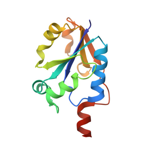
Glycerol-3-phosphate cytidylyltransferase. Structural changes induced by binding of CDP-glycerol and the role of lysine residues in catalysis
Pattridge, K.A., Weber, C.H., Friesen, J.A., Sanker, S., Kent, C., Ludwig, M.L.(2003) J Biol Chem 278: 51863-51871
- PubMed: 14506262
- DOI: https://doi.org/10.1074/jbc.M306174200
- Primary Citation of Related Structures:
1N1D - PubMed Abstract:
The bacterial enzyme, glycerol-3-phosphate cytidylyltransferase (GCT), is a model for mammalian cytidylyltransferases and is a member of a large superfamily of nucleotidyltransferases. Dimeric GCT from Bacillus subtilis displays unusual negative cooperativity in substrate binding and appears to form products only when both active sites are occupied by substrates. Here we describe a complex of GCT with the product, CDP-glycerol, in a crystal structure in which bound sulfate serves as a partial mimic of the second product, pyrophosphate. Binding of sulfate to form a pseudo-ternary complex is observed in three of the four chains constituting the asymmetric unit and is accompanied by a backbone rearrangement at Asp11 and ordering of the C-terminal helix. Comparison with the CTP complex of GCT, determined previously, reveals that in the product complex the active site closes around the glycerol phosphate moiety with a concerted motion of the segment 37-47 that includes helix B. This rearrangement allows lysines 44 and 46 to interact with the glycerol and cytosine phosphates of CDP-glycerol. Binding of CDP-glycerol also induces smaller movements of residues 92-100. Roles of lysines 44 and 46 in catalysis have been confirmed by mutagenesis of these residues to alanine, which decreases Vmax(app) and has profound effects on the Km(app) for glycerol-3-phosphate.
Organizational Affiliation:
Biophysics Research Division, University of Michigan, Ann Arbor, Michigan 48109-1055, USA.