2OQZ

The crystal structure of sortase B from B.anthracis in complex with AAEK2


Experimental Data Snapshot

  • Method: X-RAY DIFFRACTION
  • Resolution: 1.60 Å
  • R-Value Free: 0.226 
  • R-Value Work: 0.191 
  • R-Value Observed: 0.192 

wwPDB Validation   3D Report Full Report


This is version 1.3 of the entry. See complete history


Literature

Activation of inhibitors by sortase triggers irreversible modification of the active site.

Maresso, A.W.Wu, R.Kern, J.W.Zhang, R.Janik, D.Missiakas, D.M.Duban, M.E.Joachimiak, A.Schneewind, O.

(2007) J Biol Chem 282: 23129-23139

  • DOI: https://doi.org/10.1074/jbc.M701857200
  • Primary Citation of Related Structures:  
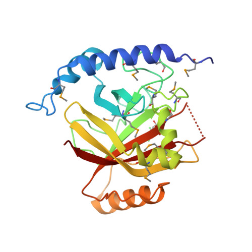
    2OQW, 2OQZ

  • PubMed Abstract: 

    Sortases anchor surface proteins to the cell wall of Gram-positive pathogens through recognition of specific motif sequences. Loss of sortase leads to large reductions in virulence, which identifies sortase as a target for the development of antibacterials. By screening 135,625 small molecules for inhibition, we report here that aryl (beta-amino)ethyl ketones inhibit sortase enzymes from staphylococci and bacilli. Inhibition of sortases occurs through an irreversible, covalent modification of their active site cysteine. Sortases specifically activate this class of molecules via beta-elimination, generating a reactive olefin intermediate that covalently modifies the cysteine thiol. Analysis of the three-dimensional structure of Bacillus anthracis sortase B with and without inhibitor provides insights into the mechanism of inhibition and reveals binding pockets that can be exploited for drug discovery.


  • Organizational Affiliation

    Department of Microbiology, University of Chicago, Chicago, Illinois 60637, USA.


Macromolecules
Find similar proteins by:  (by identity cutoff)  |  3D Structure
Entity ID: 1
MoleculeChains Sequence LengthOrganismDetailsImage
Sortase B223Bacillus anthracis str. AmesMutation(s): 11 
Gene Names: BA_4783BAS4438GBAA4783
EC: 3.4.22.51
UniProt
Find proteins for A0A6L8PZR0 (Bacillus anthracis)
Explore A0A6L8PZR0 
Go to UniProtKB:  A0A6L8PZR0
Entity Groups  
Sequence Clusters30% Identity50% Identity70% Identity90% Identity95% Identity100% Identity
UniProt GroupA0A6L8PZR0
Sequence Annotations
Expand
  • Reference Sequence
Small Molecules
Modified Residues  2 Unique
IDChains TypeFormula2D DiagramParent
CS4
Query on CS4
A
L-PEPTIDE LINKINGC12 H13 Cl2 N O3 SCYS
MSE
Query on MSE
A
L-PEPTIDE LINKINGC5 H11 N O2 SeMET
Experimental Data & Validation

Experimental Data

  • Method: X-RAY DIFFRACTION
  • Resolution: 1.60 Å
  • R-Value Free: 0.226 
  • R-Value Work: 0.191 
  • R-Value Observed: 0.192 
  • Space Group: P 1 21 1
Unit Cell:
Length ( Å )Angle ( ˚ )
a = 40.49α = 90
b = 64.964β = 105.06
c = 43.653γ = 90
Software Package:
Software NamePurpose
REFMACrefinement
HKL-2000data collection
HKL-2000data reduction
HKL-2000data scaling

Structure Validation

View Full Validation Report


Entry History 

Deposition Data

Revision History  (Full details and data files)

  • Version 1.0: 2007-06-19
    Type: Initial release
  • Version 1.1: 2008-05-01
    Changes: Version format compliance
  • Version 1.2: 2011-07-13
    Changes: Advisory, Source and taxonomy, Version format compliance
  • Version 1.3: 2018-01-24
    Changes: Advisory, Structure summary