2RHX

Crystal structure of the 3-MBT repeats from human L3MBTL1 bound to dimethyl-lysine


Experimental Data Snapshot

  • Method: X-RAY DIFFRACTION
  • Resolution: 2.10 Å
  • R-Value Free: 0.226 
  • R-Value Work: 0.196 

wwPDB Validation   3D Report Full Report


Ligand Structure Quality Assessment 


This is version 1.2 of the entry. See complete history


Literature

Structural Basis for Lower Lysine Methylation State-Specific Readout by MBT Repeats of L3MBTL1 and an Engineered PHD Finger.

Li, H.Fischle, W.Wang, W.Duncan, E.M.Liang, L.Murakami-Ishibe, S.Allis, C.D.Patel, D.J.

(2007) Mol Cell 28: 677-691

  • DOI: https://doi.org/10.1016/j.molcel.2007.10.023
  • Primary Citation of Related Structures:  
    2RHI, 2RHU, 2RHX, 2RHY, 2RHZ, 2RI2, 2RI3, 2RI5, 2RI7

  • PubMed Abstract: 

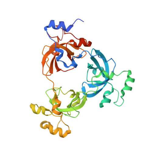
    Human L3MBTL1, which contains three malignant brain tumor (MBT) repeats, binds monomethylated and dimethylated lysines, but not trimethylated lysines, in several histone sequence contexts. In crystal structures of L3MBTL1 complexes, the monomethyl- and dimethyllysines insert into a narrow and deep cavity of aromatic residue-lined pocket 2, while a proline ring inserts into shallower pocket 1. We have also engineered a single Y to E substitution within the aromatic cage of the BPTF PHD finger, resulting in a reversal of binding preference from trimethyl- to dimethyllysine in an H3K4 sequence context. In both the "cavity insertion" (L3MBTL1) and "surface groove" (PHD finger) modes of methyllysine recognition, a carboxylate group both hydrogen bonds and ion pairs to the methylammonium proton. Our structural and binding studies of these two modules provide insights into the molecular principles governing the decoding of lysine methylation states, thereby highlighting a methylation state-specific layer of histone mark readout impacting on epigenetic regulation.


  • Organizational Affiliation

    Structural Biology Program, Memorial Sloan-Kettering Cancer Center, New York, NY 10021, USA.


Macromolecules
Find similar proteins by:  (by identity cutoff)  |  3D Structure
Entity ID: 1
MoleculeChains Sequence LengthOrganismDetailsImage
Lethal(3)malignant brain tumor-like protein345Homo sapiensMutation(s): 0 
Gene Names: L3MBTLKIAA0681L3MBT
UniProt & NIH Common Fund Data Resources
Find proteins for Q9Y468 (Homo sapiens)
Explore Q9Y468 
Go to UniProtKB:  Q9Y468
PHAROS:  Q9Y468
GTEx:  ENSG00000185513 
Entity Groups  
Sequence Clusters30% Identity50% Identity70% Identity90% Identity95% Identity100% Identity
UniProt GroupQ9Y468
Sequence Annotations
Expand
  • Reference Sequence
Experimental Data & Validation

Experimental Data

  • Method: X-RAY DIFFRACTION
  • Resolution: 2.10 Å
  • R-Value Free: 0.226 
  • R-Value Work: 0.196 
  • Space Group: P 21 21 2
Unit Cell:
Length ( Å )Angle ( ˚ )
a = 86.272α = 90
b = 93.104β = 90
c = 59.286γ = 90
Software Package:
Software NamePurpose
ADSCdata collection
MOLREPphasing
CNSrefinement
HKL-2000data reduction
HKL-2000data scaling

Structure Validation

View Full Validation Report


Ligand Structure Quality Assessment 


Entry History 

Deposition Data

Revision History  (Full details and data files)

  • Version 1.0: 2007-12-11
    Type: Initial release
  • Version 1.1: 2011-07-13
    Changes: Version format compliance
  • Version 1.2: 2023-08-30
    Changes: Data collection, Database references, Derived calculations, Refinement description