3QSC

The first crystal structure of a human telomeric G-quadruplex DNA bound to a metal-containing ligand (a copper complex)


Experimental Data Snapshot

  • Method: X-RAY DIFFRACTION
  • Resolution: 2.40 Å
  • R-Value Free: 0.234 
  • R-Value Work: 0.213 
  • R-Value Observed: 0.214 

wwPDB Validation   3D Report Full Report


Ligand Structure Quality Assessment 


This is version 1.2 of the entry. See complete history


Literature

Molecular basis of structure-activity relationships between salphen metal complexes and human telomeric DNA quadruplexes.

Campbell, N.H.Karim, N.H.Parkinson, G.N.Gunaratnam, M.Petrucci, V.Todd, A.K.Vilar, R.Neidle, S.

(2012) J Med Chem 55: 209-222

  • DOI: https://doi.org/10.1021/jm201140v
  • Primary Citation of Related Structures:  
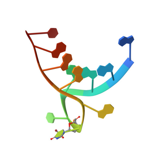
    3QSC, 3QSF

  • PubMed Abstract: 

    The first X-ray crystal structures of nickel(II) and copper(II) salphen metal complexes bound to a quadruplex DNA are presented. Two structures have been determined and show that these salphen-metal complexes bind to human telomeric quadruplexes by end-stacking, with the metal in each case almost in line with the potassium ion channel. Quadruplex and duplex DNA binding is presented for these two and other related salphen complexes, all with side-chains terminating in pyrrolidino end-groups and differing patterns of substitution on the salphen core. The crystal structures are able to provide rationalizations for the structure-activity data, and in particular for the superior quadruplex-binding of the nickel complexes compared to that of the copper-containing ones. The complexes show significant antiproliferative activity for the compounds in a panel of cancer cell lines. They also show telomerase inhibitory activity in the telomerase TRAP-LIG assay.


  • Organizational Affiliation

    CRUK Biomolecular Structure Group, The School of Pharmacy, University of London, 29-29 Brunswick Square, London WC1N 1AX, U.K.


Macromolecules

Find similar nucleic acids by:  Sequence   |   3D Structure  

Entity ID: 1
MoleculeChains LengthOrganismImage
5'-D(*AP*GP*GP*GP*TP*(BRU)P*AP*GP*GP*GP*TP*T)-3'A [auth X]12N/A
Sequence Annotations
Expand
  • Reference Sequence
Small Molecules
Ligands 2 Unique
IDChains Name / Formula / InChI Key2D Diagram3D Interactions
CUF
Query on CUF

Download Ideal Coordinates CCD File 
E [auth X][2,2'-{(4,5-difluorobenzene-1,2-diyl)bis[(nitrilo-kappaN)methylylidene]}bis{5-[2-(piperidin-1-yl)ethoxy]phenolato-kappaO}(2-)]copper (II)
C34 H38 Cu F2 N4 O4
AXRKNIOXRLONLO-OHUAMDITSA-L
K
Query on K

Download Ideal Coordinates CCD File 
B [auth X],
C [auth X],
D [auth X]
POTASSIUM ION
K
NPYPAHLBTDXSSS-UHFFFAOYSA-N
Experimental Data & Validation

Experimental Data

  • Method: X-RAY DIFFRACTION
  • Resolution: 2.40 Å
  • R-Value Free: 0.234 
  • R-Value Work: 0.213 
  • R-Value Observed: 0.214 
  • Space Group: C 2 2 2
Unit Cell:
Length ( Å )Angle ( ˚ )
a = 43.753α = 90
b = 54.786β = 90
c = 33.43γ = 90
Software Package:
Software NamePurpose
MOSFLMdata reduction
SCALAdata scaling
SHELXphasing
REFMACrefinement
PDB_EXTRACTdata extraction
ADSCdata collection
SHELXDphasing

Structure Validation

View Full Validation Report


Ligand Structure Quality Assessment 


Entry History 

Deposition Data

Revision History  (Full details and data files)

  • Version 1.0: 2011-12-07
    Type: Initial release
  • Version 1.1: 2012-04-18
    Changes: Database references
  • Version 1.2: 2023-09-13
    Changes: Data collection, Database references, Derived calculations, Refinement description