Structures of m-iodo Hoechst-DNA complexes in crystals with reduced solvent content: implications for minor groove binder drug design.
Squire, C.J., Baker, L.J., Clark, G.R., Martin, R.F., White, J.(2000) Nucleic Acids Res 28: 1252-1258
- PubMed: 10666470
- DOI: https://doi.org/10.1093/nar/28.5.1252
- Primary Citation of Related Structures:
442D, 443D, 444D, 445D, 447D, 448D, 449D - PubMed Abstract:
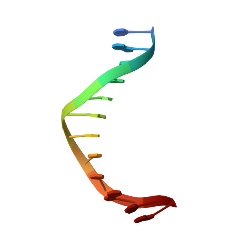
The DNA photosensitisers m-iodo Hoechst and m-iodo, p-methoxy Hoechst have been co-crystallised with the oligonucleotide d(CGCGAATTCGCG)(2)and their crystal structures determined. The crystals were then subjected to slow dehydration, which reduced their solvent contents from 40 (normal) to 30 (partially dehydrated) and then 20% (fully dehydrated) and caused a reduction in cell volume from 68,000 to 60,000 then 51,000 A(3). The dehydration resulted in a dramatic enhancement of diffraction resolution from approximately 2.6 to beyond 1.5 A. Crystal structures have also been determined for the partially and fully dehydrated states. The fully dehydrated crystals consist of an infinite polymeric network, in which neighbouring dodecamer duplexes are crosslinked through phosphate oxygens via direct bonding to bridging magnesium cations. This unique three-dimensional structure for DNA is described in detail in the following companion paper. The present paper details evidence from the sequence of crystal structures that the DNA is able to breathe locally, allowing the ligand to leave the minor groove, re-orient in the surrounding solvent medium and then re-enter the groove in a different orientation and location. The rearrangement of the minor groove binding ligands during the dehydration process mimics the binding behaviour of these ligands in solution and in vivo. We also present details of the DNA-ligand interactions that are consistent with a hydrogen atom ion mechanism for photocleavage of DNA.
Organizational Affiliation:
Chemistry Department, University of Auckland, Auckland, New Zealand.