4ZVN

Reduced quinone reductase 2 in complex with acridine orange


Experimental Data Snapshot

  • Method: X-RAY DIFFRACTION
  • Resolution: 1.87 Å
  • R-Value Free: 0.196 
  • R-Value Work: 0.160 
  • R-Value Observed: 0.162 

wwPDB Validation   3D Report Full Report


Ligand Structure Quality Assessment 


This is version 1.3 of the entry. See complete history


Literature

Binding of DNA-Intercalating Agents to Oxidized and Reduced Quinone Reductase 2.

Leung, K.K.Shilton, B.H.

(2015) Biochemistry 54: 7438-7448

  • DOI: https://doi.org/10.1021/acs.biochem.5b00884
  • Primary Citation of Related Structures:  
    4ZVK, 4ZVL, 4ZVM, 4ZVN

  • PubMed Abstract: 

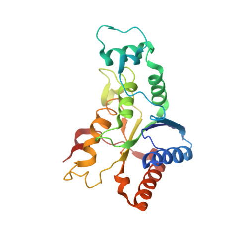
    Quinone reductase 2 (NQO2) is an enzyme that might have intracellular signaling functions. NQO2 can exist in either an oxidized state or a reduced state, and binding of compounds to one or both of these states inhibits enzymatic activity and could also affect intracellular signaling. A wide range of planar aromatic compounds bind NQO2, and we have identified three DNA-intercalating agents [ethidium bromide, acridine orange (AO), and doxorubicin] as novel nanomolar inhibitors of NQO2. Ethidium and AO, which carry a positive charge in their aromatic ring systems, bound reduced NQO2 with an affinity 50-fold higher than that of oxidized NQO2, while doxorubicin bound only oxidized NQO2. Crystallographic analyses of oxidized NQO2 in complex with the inhibitors indicated that the inhibitors were situated deep in the active site. The aromatic faces were sandwiched between the isoalloxazine ring of FAD and the phenyl ring of F178, with their edges making direct contact with residues lining the active site. In reduced NQO2, ethidium and AO occupied a more peripheral position in the active site, allowing several water molecules to interact with the polar end of the negatively charged isoalloxazine ring. We also showed that AO inhibited NQO2 at a nontoxic concentration in cells while ethidium was less effective at inhibiting NQO2 in cells. Together, this study shows that reduced NQO2 has structural and electrostatic properties that yield a preference for binding of planar, aromatic, and positively charged molecules that can also function as DNA-intercalating agents.


  • Organizational Affiliation

    Department of Biochemistry, University of Western Ontario , 1151 Richmond Street, London, Ontario, Canada N6A 5C1.


Macromolecules
Find similar proteins by:  (by identity cutoff)  |  3D Structure
Entity ID: 1
MoleculeChains Sequence LengthOrganismDetailsImage
Ribosyldihydronicotinamide dehydrogenase [quinone]
A, B
230Homo sapiensMutation(s): 0 
Gene Names: NQO2NMOR2
EC: 1.10.99.2
UniProt & NIH Common Fund Data Resources
Find proteins for P16083 (Homo sapiens)
Explore P16083 
Go to UniProtKB:  P16083
PHAROS:  P16083
GTEx:  ENSG00000124588 
Entity Groups  
Sequence Clusters30% Identity50% Identity70% Identity90% Identity95% Identity100% Identity
UniProt GroupP16083
Sequence Annotations
Expand
  • Reference Sequence
Small Molecules
Binding Affinity Annotations 
IDSourceBinding Affinity
AO Binding MOAD:  4ZVN Kd: 0.36 (nM) from 1 assay(s)
Experimental Data & Validation

Experimental Data

  • Method: X-RAY DIFFRACTION
  • Resolution: 1.87 Å
  • R-Value Free: 0.196 
  • R-Value Work: 0.160 
  • R-Value Observed: 0.162 
  • Space Group: P 21 21 21
Unit Cell:
Length ( Å )Angle ( ˚ )
a = 56.64α = 90
b = 82.86β = 90
c = 106.23γ = 90
Software Package:
Software NamePurpose
PHENIXrefinement
SCALAdata scaling
PHASERphasing
PDB_EXTRACTdata extraction
MOSFLMdata reduction

Structure Validation

View Full Validation Report


Ligand Structure Quality Assessment 


Entry History 

Deposition Data

Revision History  (Full details and data files)

  • Version 1.0: 2016-03-30
    Type: Initial release
  • Version 1.1: 2016-06-08
    Changes: Database references
  • Version 1.2: 2017-11-22
    Changes: Database references, Derived calculations, Refinement description
  • Version 1.3: 2023-09-27
    Changes: Data collection, Database references, Derived calculations, Refinement description