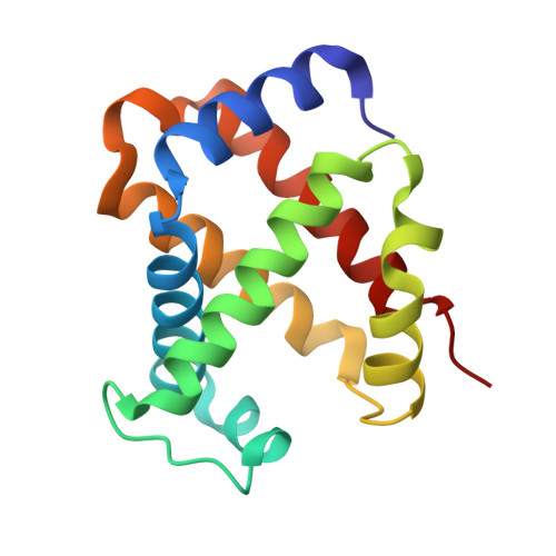
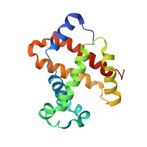
4N7P

Capturing the haemoglobin allosteric transition in a single crystal form; Crystal structure of half-liganded human haemoglobin without phosphate at 2.8 A resolution.


Experimental Data Snapshot

  • Method: X-RAY DIFFRACTION
  • Resolution: 2.81 Å
  • R-Value Free: 0.287 
  • R-Value Work: 0.259 

wwPDB Validation   3D Report Full Report


Ligand Structure Quality Assessment 


This is version 1.3 of the entry. See complete history


Literature

Capturing the hemoglobin allosteric transition in a single crystal form

Shibayama, N.Sugiyama, K.Tame, J.R.Park, S.Y.

(2014) J Am Chem Soc 136: 5097-5105

  • DOI: https://doi.org/10.1021/ja500380e
  • Primary Citation of Related Structures:  
    4N7N, 4N7O, 4N7P

  • PubMed Abstract: 

    Allostery in many oligomeric proteins has been postulated to occur via a ligand-binding-driven conformational transition from the tense (T) to relaxed (R) state, largely on the basis of the knowledge of the structure and function of hemoglobin, the most thoroughly studied of all allosteric proteins. However, a growing body of evidence suggests that hemoglobin possesses a variety of intermediates between the two end states. As such intermediate forms coexist with the end states in dynamic equilibrium and cannot be individually characterized by conventional techniques, very little is known about their properties and functions. Here we present complete structural and functional snapshots of nine equilibrium conformers of human hemoglobin in the half-liganded and fully liganded states by using a novel combination of X-ray diffraction analysis and microspectrophotometric O2 equilibrium measurements on three isomorphous crystals, each capturing three distinct equilibrium conformers. Notably, the conformational set of this crystal form varies according to shifts in the allosteric equilibrium, reflecting the differences in hemoglobin ligation state and crystallization solution conditions. We find that nine snapshot structures cover the complete conformational space of hemoglobin, ranging from T to R2 (the second relaxed quaternary structure) through R, with various relaxed intermediate forms between R and R2. Moreover, we find a previously unidentified intermediate conformer, between T and R, with an intermediate O2 affinity, sought by many research groups over a period of decades. These findings reveal a comprehensive picture of the equilibrium conformers and transition pathway for human hemoglobin.


  • Organizational Affiliation

    Division of Biophysics, Department of Physiology, Jichi Medical University , 3311-1 Yakushiji, Shimotsuke, Tochigi 329-0498, Japan.


Macromolecules
Find similar proteins by:  (by identity cutoff)  |  3D Structure
Entity ID: 1
MoleculeChains Sequence LengthOrganismDetailsImage
Hemoglobin subunit alpha
A, C, E, G, I
A, C, E, G, I, K
141Homo sapiensMutation(s): 0 
UniProt & NIH Common Fund Data Resources
Find proteins for P69905 (Homo sapiens)
Explore P69905 
Go to UniProtKB:  P69905
PHAROS:  P69905
Entity Groups  
Sequence Clusters30% Identity50% Identity70% Identity90% Identity95% Identity100% Identity
UniProt GroupP69905
Sequence Annotations
Expand
  • Reference Sequence
Find similar proteins by:  (by identity cutoff)  |  3D Structure
Entity ID: 2
MoleculeChains Sequence LengthOrganismDetailsImage
Hemoglobin subunit beta
B, D, F, H, J
B, D, F, H, J, L
146Homo sapiensMutation(s): 0 
UniProt & NIH Common Fund Data Resources
Find proteins for P68871 (Homo sapiens)
Explore P68871 
Go to UniProtKB:  P68871
PHAROS:  P68871
GTEx:  ENSG00000244734 
Entity Groups  
Sequence Clusters30% Identity50% Identity70% Identity90% Identity95% Identity100% Identity
UniProt GroupP68871
Sequence Annotations
Expand
  • Reference Sequence
Small Molecules
Ligands 2 Unique
IDChains Name / Formula / InChI Key2D Diagram3D Interactions
HNI
Query on HNI

Download Ideal Coordinates CCD File 
M [auth A]
P [auth D]
Q [auth E]
T [auth H]
U [auth I]
M [auth A],
P [auth D],
Q [auth E],
T [auth H],
U [auth I],
X [auth L]
PROTOPORPHYRIN IX CONTAINING NI(II)
C34 H32 N4 Ni O4
IJROJBFZULUEEG-RGGAHWMASA-L
HEM
Query on HEM

Download Ideal Coordinates CCD File 
N [auth B]
O [auth C]
R [auth F]
S [auth G]
V [auth J]
N [auth B],
O [auth C],
R [auth F],
S [auth G],
V [auth J],
W [auth K]
PROTOPORPHYRIN IX CONTAINING FE
C34 H32 Fe N4 O4
KABFMIBPWCXCRK-RGGAHWMASA-L
Experimental Data & Validation

Experimental Data

  • Method: X-RAY DIFFRACTION
  • Resolution: 2.81 Å
  • R-Value Free: 0.287 
  • R-Value Work: 0.259 
  • Space Group: C 1 2 1
Unit Cell:
Length ( Å )Angle ( ˚ )
a = 228.331α = 90
b = 55.531β = 103.08
c = 139.681γ = 90
Software Package:
Software NamePurpose
DENZOdata reduction
SCALEPACKdata scaling
PHASERphasing
PHENIXrefinement
PDB_EXTRACTdata extraction
HKL-2000data collection
CNSrefinement

Structure Validation

View Full Validation Report


Ligand Structure Quality Assessment 


Entry History 

Deposition Data

Revision History  (Full details and data files)

  • Version 1.0: 2014-04-02
    Type: Initial release
  • Version 1.1: 2014-04-23
    Changes: Database references
  • Version 1.2: 2019-07-24
    Changes: Data collection, Derived calculations
  • Version 1.3: 2024-03-20
    Changes: Data collection, Database references, Derived calculations, Source and taxonomy, Structure summary