5VYQ

Crystal structure of the N-formyltransferase Rv3404c from mycobacterium tuberculosis in complex with YDP-Qui4N and folinic acid


Experimental Data Snapshot

  • Method: X-RAY DIFFRACTION
  • Resolution: 1.60 Å
  • R-Value Free: 0.200 
  • R-Value Work: 0.167 
  • R-Value Observed: 0.168 

wwPDB Validation   3D Report Full Report


Ligand Structure Quality Assessment 


This is version 1.4 of the entry. See complete history


Literature

Biochemical Investigation of Rv3404c from Mycobacterium tuberculosis.

Dunsirn, M.M.Thoden, J.B.Gilbert, M.Holden, H.M.

(2017) Biochemistry 56: 3818-3825

  • DOI: https://doi.org/10.1021/acs.biochem.7b00506
  • Primary Citation of Related Structures:  
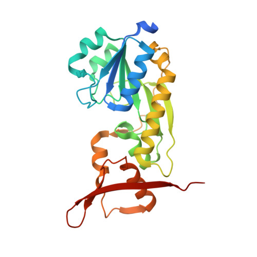
    5VYQ

  • PubMed Abstract: 

    The causative agent of tuberculosis, Mycobacterium tuberculosis, is a bacterium with a complex cell wall and a complicated life cycle. The genome of M. tuberculosis contains well over 4000 genes thought to encode proteins. One of these codes for a putative enzyme referred to as Rv3404c, which has attracted research attention as a potential virulence factor for over 12 years. Here we demonstrate that Rv3404c functions as a sugar N-formyltransferase that converts dTDP-4-amino-4,6-dideoxyglucose into dTDP-4-formamido-4,6-dideoxyglucose using N 10 -formyltetrahydrofolate as the carbon source. Kinetic analyses demonstrate that Rv3404c displays a significant catalytic efficiency of 1.1 × 10 4 M -1 s -1 . In addition, we report the X-ray structure of a ternary complex of Rv3404c solved in the presence of N 5 -formyltetrahydrofolate and dTDP-4-amino-4,6-dideoxyglucose. The final model of Rv3404c was refined to an overall R-factor of 16.8% at 1.6 Å resolution. The results described herein are especially intriguing given that there have been no published reports of N-formylated sugars associated with M. tuberculosis. The data thus provide a new avenue of research into this fascinating, yet deadly, organism that apparently has been associated with human infection since ancient times.


  • Organizational Affiliation

    Department of Biochemistry, University of Wisconsin , Madison, Wisconsin 53706, United States.


Macromolecules
Find similar proteins by:  (by identity cutoff)  |  3D Structure
Entity ID: 1
MoleculeChains Sequence LengthOrganismDetailsImage
Uncharacterized protein
A, B
255Mycobacterium tuberculosis CAS/NITR204Mutation(s): 0 
Gene Names: J113_23825
UniProt
Find proteins for P9WKZ3 (Mycobacterium tuberculosis (strain ATCC 25618 / H37Rv))
Explore P9WKZ3 
Go to UniProtKB:  P9WKZ3
Entity Groups  
Sequence Clusters30% Identity50% Identity70% Identity90% Identity95% Identity100% Identity
UniProt GroupP9WKZ3
Sequence Annotations
Expand
  • Reference Sequence
Small Molecules
Ligands 9 Unique
IDChains Name / Formula / InChI Key2D Diagram3D Interactions
0FX
Query on 0FX

Download Ideal Coordinates CCD File 
D [auth A]dTDP-4-amino-4,6-dideoxyglucose
C16 H27 N3 O14 P2
UIVJXHWSIFBBCY-LPGAPTBISA-N
FON
Query on FON

Download Ideal Coordinates CCD File 
C [auth A]N-{[4-({[(6R)-2-amino-5-formyl-4-oxo-1,4,5,6,7,8-hexahydropteridin-6-yl]methyl}amino)phenyl]carbonyl}-L-glutamic acid
C20 H23 N7 O7
VVIAGPKUTFNRDU-OLZOCXBDSA-N
TYD
Query on TYD

Download Ideal Coordinates CCD File 
N [auth B]THYMIDINE-5'-DIPHOSPHATE
C10 H16 N2 O11 P2
UJLXYODCHAELLY-XLPZGREQSA-N
EP1
Query on EP1

Download Ideal Coordinates CCD File 
J [auth A],
R [auth B]
3-[4-(2-HYDROXYETHYL)PIPERAZIN-1-YL]PROPANE-1-SULFONIC ACID
C9 H20 N2 O4 S
OWXMKDGYPWMGEB-UHFFFAOYSA-N
EDO
Query on EDO

Download Ideal Coordinates CCD File 
E [auth A],
F [auth A],
O [auth B],
P [auth B]
1,2-ETHANEDIOL
C2 H6 O2
LYCAIKOWRPUZTN-UHFFFAOYSA-N
K
Query on K

Download Ideal Coordinates CCD File 
T [auth B]POTASSIUM ION
K
NPYPAHLBTDXSSS-UHFFFAOYSA-N
CL
Query on CL

Download Ideal Coordinates CCD File 
I [auth A],
K [auth A],
Q [auth B]
CHLORIDE ION
Cl
VEXZGXHMUGYJMC-UHFFFAOYSA-M
NA
Query on NA

Download Ideal Coordinates CCD File 
G [auth A],
H [auth A],
L [auth A],
S [auth B]
SODIUM ION
Na
FKNQFGJONOIPTF-UHFFFAOYSA-N
LI
Query on LI

Download Ideal Coordinates CCD File 
M [auth A]LITHIUM ION
Li
HBBGRARXTFLTSG-UHFFFAOYSA-N
Experimental Data & Validation

Experimental Data

  • Method: X-RAY DIFFRACTION
  • Resolution: 1.60 Å
  • R-Value Free: 0.200 
  • R-Value Work: 0.167 
  • R-Value Observed: 0.168 
  • Space Group: P 21 21 21
Unit Cell:
Length ( Å )Angle ( ˚ )
a = 47.486α = 90
b = 73.018β = 90
c = 173.265γ = 90
Software Package:
Software NamePurpose
REFMACrefinement
SAINTdata reduction
SADABSdata scaling
PHASERphasing

Structure Validation

View Full Validation Report


Ligand Structure Quality Assessment 


Entry History & Funding Information

Deposition Data


Funding OrganizationLocationGrant Number
National Institutes of Health/National Institute of General Medical Sciences (NIH/NIGMS)United StatesGM115921

Revision History  (Full details and data files)

  • Version 1.0: 2017-07-12
    Type: Initial release
  • Version 1.1: 2017-08-09
    Changes: Database references
  • Version 1.2: 2017-09-20
    Changes: Author supporting evidence
  • Version 1.3: 2020-01-01
    Changes: Author supporting evidence
  • Version 1.4: 2023-10-04
    Changes: Data collection, Database references, Derived calculations, Refinement description