5ZTF

Structure of Ca2+ ATPase


Experimental Data Snapshot

  • Method: X-RAY DIFFRACTION
  • Resolution: 3.45 Å
  • R-Value Free: 0.307 
  • R-Value Work: 0.246 
  • R-Value Observed: 0.249 

wwPDB Validation   3D Report Full Report


Ligand Structure Quality Assessment 


This is version 1.1 of the entry. See complete history


Literature

Structural Basis of Sarco/Endoplasmic Reticulum Ca2+-ATPase 2b Regulation via Transmembrane Helix Interplay.

Inoue, M.Sakuta, N.Watanabe, S.Zhang, Y.Yoshikaie, K.Tanaka, Y.Ushioda, R.Kato, Y.Takagi, J.Tsukazaki, T.Nagata, K.Inaba, K.

(2019) Cell Rep 27: 1221-1230.e3

  • DOI: https://doi.org/10.1016/j.celrep.2019.03.106
  • Primary Citation of Related Structures:  
    5ZTF, 6JJU

  • PubMed Abstract: 

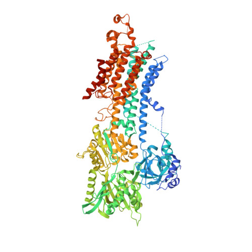
    Sarco/endoplasmic reticulum (ER) Ca 2+ -ATPase 2b (SERCA2b) is a ubiquitously expressed membrane protein that facilitates Ca 2+ uptake from the cytosol to the ER. SERCA2b includes a characteristic 11 th transmembrane helix (TM11) followed by a luminal tail, but the structural basis of SERCA regulation by these C-terminal segments remains unclear. Here, we determined the crystal structures of SERCA2b and its C-terminal splicing variant SERCA2a, both in the E1-2Ca 2+ -adenylyl methylenediphosphonate (AMPPCP) state. Despite discrepancies with the previously reported structural model of SERCA2b, TM11 was found to be located adjacent to TM10 and to interact weakly with a part of the L8/9 loop and the N-terminal end of TM10, thereby inhibiting the SERCA2b catalytic cycle. Accordingly, mutational disruption of the interactions between TM11 and its neighboring residues caused SERCA2b to display SERCA2a-like ATPase activity. We propose that TM11 serves as a key modulator of SERCA2b activity by fine-tuning the intramolecular interactions with other transmembrane regions.


  • Organizational Affiliation

    Institute of Multidisciplinary Research for Advanced Materials, Tohoku University, Sendai 980-8577, Japan; Core Research for Evolutional Science and Technology (CREST), Japan Science and Technology Agency, Saitama, Kawaguchi, Japan.


Macromolecules
Find similar proteins by:  (by identity cutoff)  |  3D Structure
Entity ID: 1
MoleculeChains Sequence LengthOrganismDetailsImage
Sarcoplasmic/endoplasmic reticulum calcium ATPase 21,070Homo sapiensMutation(s): 0 
Gene Names: ATP2A2ATP2B
EC: 3.6.3.8
Membrane Entity: Yes 
UniProt & NIH Common Fund Data Resources
Find proteins for P16615 (Homo sapiens)
Explore P16615 
Go to UniProtKB:  P16615
PHAROS:  P16615
GTEx:  ENSG00000174437 
Entity Groups  
Sequence Clusters30% Identity50% Identity70% Identity90% Identity95% Identity100% Identity
UniProt GroupP16615
Sequence Annotations
Expand
  • Reference Sequence
Experimental Data & Validation

Experimental Data

  • Method: X-RAY DIFFRACTION
  • Resolution: 3.45 Å
  • R-Value Free: 0.307 
  • R-Value Work: 0.246 
  • R-Value Observed: 0.249 
  • Space Group: P 21 21 2
Unit Cell:
Length ( Å )Angle ( ˚ )
a = 165.079α = 90
b = 84.053β = 90
c = 118.832γ = 90
Software Package:
Software NamePurpose
PHENIXrefinement
XDSdata reduction
Aimlessdata scaling
MOLREPphasing

Structure Validation

View Full Validation Report


Ligand Structure Quality Assessment 


Entry History & Funding Information

Deposition Data


Funding OrganizationLocationGrant Number
Japan Science and TechnologyJapanJPMJCR13M6

Revision History  (Full details and data files)

  • Version 1.0: 2019-05-22
    Type: Initial release
  • Version 1.1: 2023-11-22
    Changes: Data collection, Database references, Derived calculations, Refinement description