6HSN

Crystal structure of the ternary complex of GephE-ADP-GABA(A) receptor derived peptide


Experimental Data Snapshot

  • Method: X-RAY DIFFRACTION
  • Resolution: 1.55 Å
  • R-Value Free: 0.196 
  • R-Value Work: 0.159 
  • R-Value Observed: 0.161 

wwPDB Validation   3D Report Full Report


Ligand Structure Quality Assessment 


This is version 1.4 of the entry. See complete history


Literature

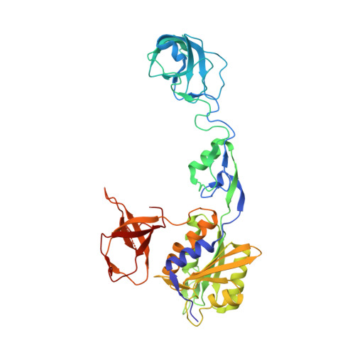
Elucidating the Molecular Basis for Inhibitory Neurotransmission Regulation by Artemisinins.

Kasaragod, V.B.Hausrat, T.J.Schaefer, N.Kuhn, M.Christensen, N.R.Tessmer, I.Maric, H.M.Madsen, K.L.Sotriffer, C.Villmann, C.Kneussel, M.Schindelin, H.

(2019) Neuron 101: 673-689.e11

  • DOI: https://doi.org/10.1016/j.neuron.2019.01.001
  • Primary Citation of Related Structures:  
    6FGC, 6FGD, 6HSN, 6HSO

  • PubMed Abstract: 

    The frontline anti-malarial drug artemisinin and its derivatives have also been implicated in modulating multiple mammalian cellular pathways, including the recent identification of targeting γ-aminobutyric acid type A receptor (GABA A R) signaling in the pancreas. Their molecular mechanism of action, however, remains elusive. Here, we present crystal structures of gephyrin, the central organizer at inhibitory postsynapses, in complex with artesunate and artemether at 1.5-Å resolution. These artemisinins target the universal inhibitory neurotransmitter receptor-binding epitope of gephyrin, thus inhibiting critical interactions between gephyrin and glycine receptors (GlyRs) as well as GABA A Rs. Electrophysiological recordings reveal a significant inhibition of gephyrin-mediated neurotransmission by artemisinins. Furthermore, clustering analyses in primary neurons demonstrate a rapid inhibition and a time-dependent regulation of gephyrin and GABA A R cluster parameters. Our data not only provide a comprehensive model for artemisinin-mediated modulation of inhibitory neurotransmission but also establish artemisinins as potential lead compounds to pharmacologically interfere with this process.


  • Organizational Affiliation

    Institute of Structural Biology, Rudolf Virchow Center for Experimental Biomedicine, University of Würzburg, Josef-Schneider-Str. 2, 97080 Würzburg, Germany.


Macromolecules
Find similar proteins by:  (by identity cutoff)  |  3D Structure
Entity ID: 1
MoleculeChains Sequence LengthOrganismDetailsImage
Gephyrin419Rattus norvegicusMutation(s): 0 
Gene Names: GphnGph
EC: 2.7.7.75 (PDB Primary Data), 2.10.1.1 (PDB Primary Data)
UniProt
Find proteins for Q03555 (Rattus norvegicus)
Explore Q03555 
Go to UniProtKB:  Q03555
Entity Groups  
Sequence Clusters30% Identity50% Identity70% Identity90% Identity95% Identity100% Identity
UniProt GroupQ03555
Sequence Annotations
Expand
  • Reference Sequence

Find similar proteins by:  Sequence   |   3D Structure  

Entity ID: 2
MoleculeChains Sequence LengthOrganismDetailsImage
Gamma-aminobutyric acid receptor subunit alpha-3B [auth D],
C [auth E]
10Rattus norvegicusMutation(s): 0 
UniProt
Find proteins for P20236 (Rattus norvegicus)
Explore P20236 
Go to UniProtKB:  P20236
Entity Groups  
Sequence Clusters30% Identity50% Identity70% Identity90% Identity95% Identity100% Identity
UniProt GroupP20236
Sequence Annotations
Expand
  • Reference Sequence
Small Molecules
Ligands 7 Unique
IDChains Name / Formula / InChI Key2D Diagram3D Interactions
ADP
Query on ADP

Download Ideal Coordinates CCD File 
D [auth A]ADENOSINE-5'-DIPHOSPHATE
C10 H15 N5 O10 P2
XTWYTFMLZFPYCI-KQYNXXCUSA-N
3F8
Query on 3F8

Download Ideal Coordinates CCD File 
T [auth D]1,1'-[ethane-1,2-diylbis(oxyethane-2,1-diyl)]bis(1H-pyrrole-2,5-dione)
C14 H16 N2 O6
FERLGYOHRKHQJP-UHFFFAOYSA-N
MPD
Query on MPD

Download Ideal Coordinates CCD File 
G [auth A],
H [auth A],
I [auth A]
(4S)-2-METHYL-2,4-PENTANEDIOL
C6 H14 O2
SVTBMSDMJJWYQN-YFKPBYRVSA-N
PO4
Query on PO4

Download Ideal Coordinates CCD File 
R [auth A]PHOSPHATE ION
O4 P
NBIIXXVUZAFLBC-UHFFFAOYSA-K
ACT
Query on ACT

Download Ideal Coordinates CCD File 
J [auth A]
K [auth A]
L [auth A]
M [auth A]
N [auth A]
J [auth A],
K [auth A],
L [auth A],
M [auth A],
N [auth A],
O [auth A],
P [auth A],
Q [auth A]
ACETATE ION
C2 H3 O2
QTBSBXVTEAMEQO-UHFFFAOYSA-M
MG
Query on MG

Download Ideal Coordinates CCD File 
E [auth A],
F [auth A]
MAGNESIUM ION
Mg
JLVVSXFLKOJNIY-UHFFFAOYSA-N
NA
Query on NA

Download Ideal Coordinates CCD File 
S [auth A]SODIUM ION
Na
FKNQFGJONOIPTF-UHFFFAOYSA-N
Experimental Data & Validation

Experimental Data

  • Method: X-RAY DIFFRACTION
  • Resolution: 1.55 Å
  • R-Value Free: 0.196 
  • R-Value Work: 0.159 
  • R-Value Observed: 0.161 
  • Space Group: I 2 2 2
Unit Cell:
Length ( Å )Angle ( ˚ )
a = 86.962α = 90
b = 99.217β = 90
c = 111.608γ = 90
Software Package:
Software NamePurpose
REFMACrefinement
XDSdata reduction
Aimlessdata scaling
PHASERphasing

Structure Validation

View Full Validation Report


Ligand Structure Quality Assessment 


Entry History & Funding Information

Deposition Data


Funding OrganizationLocationGrant Number
German Research FoundationGermanySCHI 425/ 8-2

Revision History  (Full details and data files)

  • Version 1.0: 2019-01-16
    Type: Initial release
  • Version 1.1: 2019-01-30
    Changes: Data collection, Derived calculations
  • Version 1.2: 2019-02-13
    Changes: Data collection, Database references
  • Version 1.3: 2019-03-06
    Changes: Data collection, Database references
  • Version 1.4: 2024-01-24
    Changes: Data collection, Database references, Derived calculations, Refinement description