6PWN

MscS Nanodisc with N-terminal His-Tag


Experimental Data Snapshot

  • Method: ELECTRON MICROSCOPY
  • Resolution: 3.10 Å
  • Aggregation State: PARTICLE 
  • Reconstruction Method: SINGLE PARTICLE 

wwPDB Validation   3D Report Full Report


This is version 1.1 of the entry. See complete history


Literature

Molecular basis of force-from-lipids gating in the mechanosensitive channel MscS.

Reddy, B.Bavi, N.Lu, A.Park, Y.Perozo, E.

(2019) Elife 8

  • DOI: https://doi.org/10.7554/eLife.50486
  • Primary Citation of Related Structures:  
    6PWN, 6PWO, 6PWP

  • PubMed Abstract: 

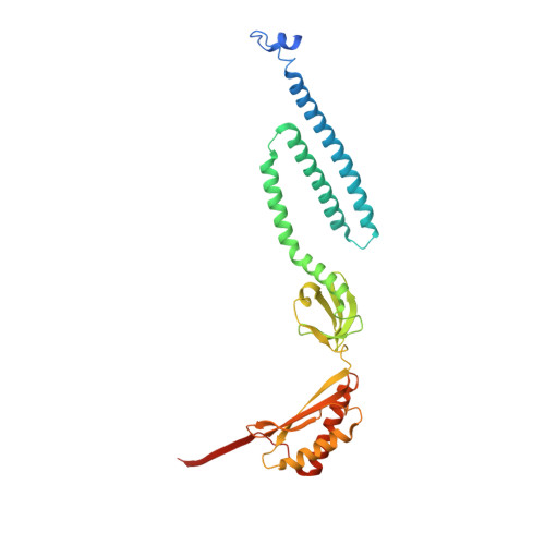
    Prokaryotic mechanosensitive (MS) channels open by sensing the physical state of the membrane. As such, lipid-protein interactions represent the defining molecular process underlying mechanotransduction. Here, we describe cryo-electron microscopy (cryo-EM) structures of the E. coli small-conductance mechanosensitive channel (MscS) in nanodiscs (ND). They reveal a novel membrane-anchoring fold that plays a significant role in channel activation and establish a new location for the lipid bilayer, shifted ~14 Å from previous consensus placements. Two types of lipid densities are explicitly observed. A phospholipid that 'hooks' the top of each TM2-TM3 hairpin and likely plays a role in force sensing, and a bundle of acyl chains occluding the permeation path above the L105 cuff. These observations reshape our understanding of force-from-lipids gating in MscS and highlight the key role of allosteric interactions between TM segments and phospholipids bound to key dynamic components of the channel.


  • Organizational Affiliation

    Department of Biochemistry and Molecular Biology, The University of Chicago, Chicago, United States.


Macromolecules
Find similar proteins by:  (by identity cutoff)  |  3D Structure
Entity ID: 1
MoleculeChains Sequence LengthOrganismDetailsImage
Small-conductance mechanosensitive channel
A, B, C, D, E
A, B, C, D, E, F, G
306Escherichia coli K-12Mutation(s): 0 
Gene Names: mscSyggBb2924JW2891
Membrane Entity: Yes 
UniProt
Find proteins for P0C0S1 (Escherichia coli (strain K12))
Explore P0C0S1 
Go to UniProtKB:  P0C0S1
Entity Groups  
Sequence Clusters30% Identity50% Identity70% Identity90% Identity95% Identity100% Identity
UniProt GroupP0C0S1
Sequence Annotations
Expand
  • Reference Sequence
Small Molecules
Ligands 2 Unique
IDChains Name / Formula / InChI Key2D Diagram3D Interactions
POV
Query on POV

Download Ideal Coordinates CCD File 
H [auth A]
J [auth B]
L [auth C]
O [auth D]
P [auth E]
H [auth A],
J [auth B],
L [auth C],
O [auth D],
P [auth E],
R [auth F],
U [auth G]
(2S)-3-(hexadecanoyloxy)-2-[(9Z)-octadec-9-enoyloxy]propyl 2-(trimethylammonio)ethyl phosphate
C42 H82 N O8 P
WTJKGGKOPKCXLL-PFDVCBLKSA-N
R16
Query on R16

Download Ideal Coordinates CCD File 
I [auth A]
K [auth B]
M [auth C]
N [auth C]
Q [auth E]
I [auth A],
K [auth B],
M [auth C],
N [auth C],
Q [auth E],
S [auth F],
T [auth F]
HEXADECANE
C16 H34
DCAYPVUWAIABOU-UHFFFAOYSA-N
Experimental Data & Validation

Experimental Data

  • Method: ELECTRON MICROSCOPY
  • Resolution: 3.10 Å
  • Aggregation State: PARTICLE 
  • Reconstruction Method: SINGLE PARTICLE 
EM Software:
TaskSoftware PackageVersion
RECONSTRUCTIONRELION3.0.6

Structure Validation

View Full Validation Report


Entry History & Funding Information

Deposition Data


Funding OrganizationLocationGrant Number
National Institutes of Health/National Human Genome Research Institute (NIH/NHGRI)United StatesR01 GM133191

Revision History  (Full details and data files)

  • Version 1.0: 2020-01-08
    Type: Initial release
  • Version 1.1: 2024-03-20
    Changes: Data collection, Database references