6S7R

Non-square conformations of KtrA R16A mutant rings with bound ADP


Experimental Data Snapshot

  • Method: X-RAY DIFFRACTION
  • Resolution: 3.73 Å
  • R-Value Free: 0.300 
  • R-Value Work: 0.246 
  • R-Value Observed: 0.251 

wwPDB Validation   3D Report Full Report


Ligand Structure Quality Assessment 


This is version 1.1 of the entry. See complete history


Literature

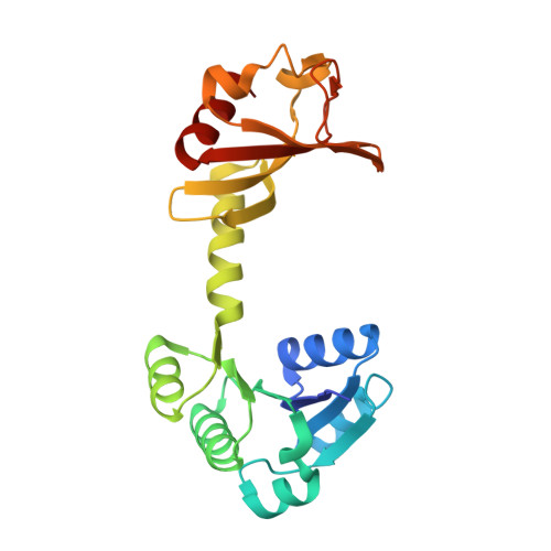
Activation of a nucleotide-dependent RCK domain requires binding of a cation cofactor to a conserved site.

Teixeira-Duarte, C.M.Fonseca, F.Morais Cabral, J.H.

(2019) Elife 8

  • DOI: https://doi.org/10.7554/eLife.50661
  • Primary Citation of Related Structures:  
    6S2J, 6S5B, 6S5C, 6S5D, 6S5E, 6S5G, 6S5N, 6S5O, 6S7R

  • PubMed Abstract: 

    RCK domains regulate the activity of K + channels and transporters in eukaryotic and prokaryotic organisms by responding to ions or nucleotides. The mechanisms of RCK activation by Ca 2+ in the eukaryotic BK and bacterial MthK K + channels are well understood. However, the molecular details of activation in nucleotide-dependent RCK domains are not clear. Through a functional and structural analysis of the mechanism of ATP activation in KtrA, a RCK domain from the B. subtilis KtrAB cation channel, we have found that activation by nucleotide requires binding of cations to an intra-dimer interface site in the RCK dimer. In particular, divalent cations are coordinated by the γ-phosphates of bound-ATP, tethering the two subunits and stabilizing the active state conformation. Strikingly, the binding site residues are highly conserved in many different nucleotide-dependent RCK domains, indicating that divalent cations are a general cofactor in the regulatory mechanism of many nucleotide-dependent RCK domains.


  • Organizational Affiliation

    Instituto de Investigação e Inovação em Saúde (i3S), Universidade do Porto, Porto, Portugal.


Macromolecules
Find similar proteins by:  (by identity cutoff)  |  3D Structure
Entity ID: 1
MoleculeChains Sequence LengthOrganismDetailsImage
Ktr system potassium uptake protein A
A, B, C, D, E
A, B, C, D, E, F, G, H, I, J, K, L, M, N, O, P
222Bacillus subtilis subsp. subtilis str. 168Mutation(s): 1 
Gene Names: ktrAyuaABSU31090
UniProt
Find proteins for O32080 (Bacillus subtilis (strain 168))
Explore O32080 
Go to UniProtKB:  O32080
Entity Groups  
Sequence Clusters30% Identity50% Identity70% Identity90% Identity95% Identity100% Identity
UniProt GroupO32080
Sequence Annotations
Expand
  • Reference Sequence
Small Molecules
Ligands 1 Unique
IDChains Name / Formula / InChI Key2D Diagram3D Interactions
ADP (Subject of Investigation/LOI)
Query on ADP

Download Ideal Coordinates CCD File 
AA [auth K]
BA [auth L]
CA [auth M]
DA [auth N]
EA [auth O]
AA [auth K],
BA [auth L],
CA [auth M],
DA [auth N],
EA [auth O],
FA [auth P],
Q [auth A],
R [auth B],
S [auth C],
T [auth D],
U [auth E],
V [auth F],
W [auth G],
X [auth H],
Y [auth I],
Z [auth J]
ADENOSINE-5'-DIPHOSPHATE
C10 H15 N5 O10 P2
XTWYTFMLZFPYCI-KQYNXXCUSA-N
Experimental Data & Validation

Experimental Data

  • Method: X-RAY DIFFRACTION
  • Resolution: 3.73 Å
  • R-Value Free: 0.300 
  • R-Value Work: 0.246 
  • R-Value Observed: 0.251 
  • Space Group: P 1
Unit Cell:
Length ( Å )Angle ( ˚ )
a = 92.967α = 90.42
b = 97.874β = 97.1
c = 144.561γ = 110.27
Software Package:
Software NamePurpose
PHENIXrefinement
XDSdata reduction
XDSdata scaling
PHASERphasing

Structure Validation

View Full Validation Report


Ligand Structure Quality Assessment 


Entry History & Funding Information

Deposition Data


Funding OrganizationLocationGrant Number
Fundacao para a Ciencia e a TecnologiaPortugalPTDC/BIA-BQM/29863/2017
Fundacao para a Ciencia e a TecnologiaPortugalPOCI-01-0145-FEDER-007274

Revision History  (Full details and data files)

  • Version 1.0: 2020-01-08
    Type: Initial release
  • Version 1.1: 2024-01-24
    Changes: Advisory, Data collection, Database references, Refinement description